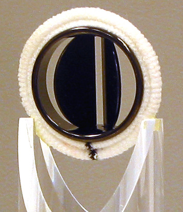
Figure 9. St. Jude bileaflet prosthesis developed in 1976.
Heart Valves (1968 and beyond)
Initial development in the field of prosthetic heart valves involved the search for biologically compatible materials and hemologically tolerant designs. At that time, as there was no satisfactory mechanism to scientifically achieve this goal, the trial and error method was used and much of this early work was performed at the University of Minnesota. The development of prosthetic heart valves became the purview of several cardiovascular surgeons who often collaborated with engineers. To distinguish one valve from the others, each prosthesis often became identified and named after its surgeon developer [9]. It is notable that Lillehei and his colleagues developed four different valves: 1) a non-tilting disc valve called the Lillehei-Nakib Toroidal Valve in 1967; 2) two tilting disc valves, the Lillehei-Cruz-Kaster in 1963 and the Lillehei-Kaster in 1970; and 3) a bileaflet valve, the Lillehei-Kalke in 1965.
The St. Jude bileaflet valve was primarily designed by Chris Posis, an industrial engineer who approached Demetre Nicoloff, MD, a cardiovascular surgeon at the University of Minnesota. This valve had floating hinges located near the central axis of the rigid housing as well as an opening to the outer edge of each leaflet, leaving a small central opening (Figure 9) [10]. Dr. Nicoloff first implanted this valve in 1976, and it provided the foundation for the beginning of St. Jude Medical as a significant biomedical device company.
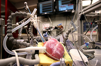
Figure 10. A functioning isolated heart in the Visible Heart Laboratory.
Visible Heart Laboratory (1997)
In 1997, Dr. Paul Iaizzo and his coworkers began working on large mammalian isolated heart models (Figure 10), and thus the Visible Heart® laboratory was created in collaboration with Medtronic, Inc. Today, this lab is a premiere place to perform translational systems physiology research which ranges from cellular and tissue studies to organ and whole body investigations. The Visible Heart® lab embodies a creative atmosphere which is energized by some of the best and brightest students at the University. Our lab staff has over 100 years of collective research experience and functions as a highly efficient and productive team.
|


