University of Minnesota
http://www.umn.edu/
612-625-5000
What is Echocardiography?
Echocardiography is a diagnostic tool that uses ultrasound to image the heart. It is useful for assessing cardiac structure and function. It can be used to obtain quantitative measurements like valve, chamber and vessel sizes and quantitative assessment of ventricular systolic and diastolic function. Ultrasound is free from radiation and is safe to use in fetal, neonatal, pediatric, and adult patients.
Types
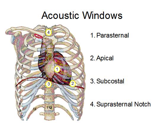
Transthoracic echocardiography (TTE) is the most common type. The images are obtained by placing the probe in the chest, and are complicated by patient size, bone, soft tissue, and lung tissue interactions. As a result, the ultrasound beams from the transducer head must be aimed through narrow acoustic windows in the thorax (Figure 1). TTE It is performed with a phased array probe that creates a wedge-shaped image with the closest structure to the probe located at the tip of the triangle.
Figure 1: Transthoracic echocardiography obtains cardiac images by placing the transducer head in contact with the thorax at one of the following locations, or acoustic windows: parasternal, apical, subcostal, and the suprasternal notch.
TTE. Four chamber view. Probe is placed in the apical position. Notice that the apex of the heart is closest to the probe (superior part of the screen) and the atria are farthest away (bottom of the screen) as opposed to TEE.
Transesophageal echocardiography (TEE) involves the placement of an echocardiography transducer into the esophagus or the stomach of an anesthetized or sedated patient. This placement eliminates the need to use acoustic windows, minimizes interference from the lungs, and increases the resolution of the images obtained, due to the closer proximity of the transducer to the heart. TEE is useful for intraoperative monitoring of anesthetized patients and is used for patients in which TTE did not produce quality images. Similar to TTE a wedge image is created, in this case the closes structure to the probe is the left atrium and the farthest the apex of the heart.
TEE. ME – four chamber view. Notice that the apex of the heart is farthest from the probe (bottom part of the screen) and the atria are closest (superior part of the screen) as opposed to TTE.
Intracardiac echocardiography (ICE) provides better resolution than either TTE or TEE. The ICE catheter is advanced through a standard femoral venous introducer to the right atrium. By placing the transducer directly into the heart, higher frequencies of sound waves can be used, creating excellent spatial resolution.