Your Heart and the Cardiovascular System
Your Heart is just one of the components of your body’s cardiovascular system. This system can be broken
down into several components which includes: blood, blood vessels, coronary circulation, and the heart.
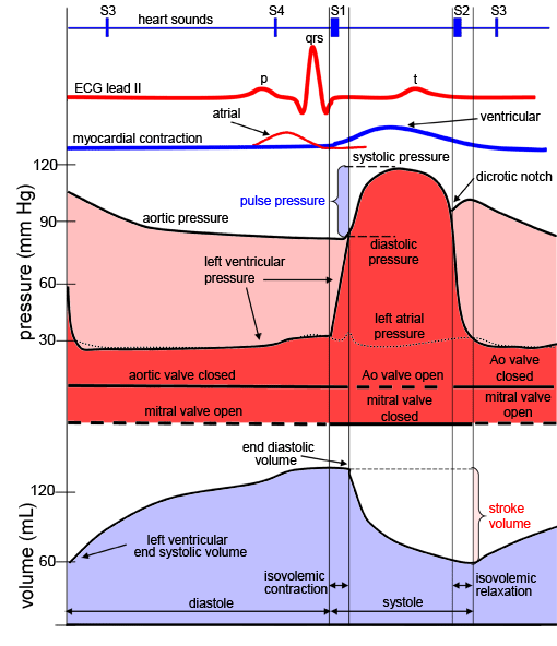
During a single cardiac cycle, the atria and ventricles do not beat simultaneously; the atrial
contraction occurs prior to ventricular contraction. This timing delay allows for proper filling of
all four chambers of the heart. Recall that the left and right heart pumps function in parallel.
The diastolic phase of the cardiac cycle begins with the opening of the tricuspid and mitral valves
(atrioventricular valves). The atrioventricular valves open when the pressures in the ventricles
fall below those in the atria. This can be observed in here for the left heart, in which the mitral
valve opens when the left ventricular pressure falls below the left atrial pressure. At this
moment, passive filling of the ventricle begins. In other words, blood that has accumulated in the
atria behind the closed atrioventricular valves passes rapidly into the ventricles, and this causes
an initial drop in the atrial pressures. Later, pressures in all four chambers rise together as the
atria and ventricles continue to passively fill in unison with blood returning to the heart through
the veins (pulmonary veins to the left atrium, and the superior and inferior vena cava to the right
atrium).
Contractions of the atria are initiated near the end of ventricular diastole, which is initiated
by depolarization of the atrial myocardial cells (sinoatrial node). Atrial depolarization is
elicited at the P wave of the electrocardiogram (ECG lead II trace). The excitation and subsequent
development of tension and shortening of atrial cells cause atrial pressures to rise. Active atrial
contraction forces additional volumes of blood into the ventricles (often referred to as "atrial
kick"). The atrial kick contributes a significant volume of blood toward ventricular preload
(approximately 20%). At normal heart rates, the atrial contractions are considered essential for
adequate ventricular filling. As heart rates increase, atrial filling becomes increasingly
important for ventricular filling because the time interval between contractions for passive filling
becomes progressively shorter. Atrial fibrillation and/or asynchronized atrial-ventricular
contractions can result in minimal contribution to preload, via atrial contraction. Throughout
diastole, atrial and ventricular pressures are nearly identical due to the open atrioventricular
values which offer little or no resistance to blood flow. It should also be noted that contraction
and movement of blood out of the atrial appendage (auricle) can be an additional source for
increased blood volume.
Ventricular systole begins when the excitation passes from the right atrium through the
atrioventricular node, and through the remainder of the conduction system (His bundle and left and
right bundle branches) to cause ventricular myocardial activation. This depolarization of
ventricular cells underlies the QRS complex within the ECG. As the ventricular cells contract,
intraventricular pressures increase above those in the atria, and the atrioventricular valves
abruptly close. Closure of the atrioventricular valves results in the first heart sound, S1. As
pressures in the ventricles continue to rise together in a normally functioning heart, they
eventually reach a critical threshold pressure at which the semilunar valves (pulmonary valve and
aortic valve) open.
|


