|
The heart lies in the center of the thoracic cavity (see also web Anatomy Tutorial section) and
is suspended by its attachment to the great vessels within a fibrous sac known as the pericardium;
note that humans have relatively thick walled pericardiums compared to those of the commonly studied
large mammalian cardiovascular models (i.e., dog, pig or sheep). A small amount of fluid is present
within the sac (pericardial fluid) which lubricates the surface of the heart and allows it to move
freely during function (contraction and relaxation). The pericardial sac extends upwards enclosing
the great vessels.
|
The pathway of blood flow through the chambers of the heart is indicated in Figure 4. Recall
that venous blood returns from the systemic organs to the right atrium via the superior and inferior
venae cavae. It next passes through the tricuspid valve into the right ventricles, and from there
is pumped through the pulmonary valve into the pulmonary artery. After passing through the
pulmonary capillary beds, the oxygenated pulmonary venous blood returns to the left atrium through
the pulmonary veins. The flow of blood then passes through the mitral valve into the left
ventricle, and is pumped through the aortic valve into the aorta.
In general, the gross anatomy of the right heart pump is considerably different from that of the
left heart pump, yet the pumping principles of each are primarily the same. The ventricles are
closed chambers surrounded by muscular walls, and the valves are structurally designed to allow flow
in only one direction. The cardiac valves passively open and close in response to the direction of
the pressure gradient across them.
|
|
|
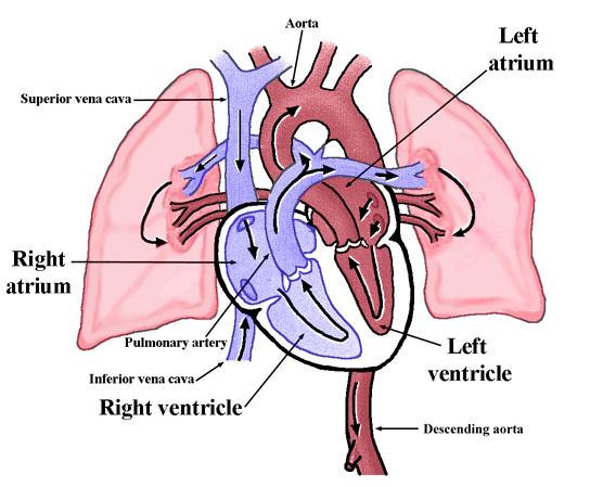
Figure 4. Pathway of blood flow through the heart and lungs. Note that the pulmonary artery (trunk)
branches into left and right pulmonary arteries. There are commonly four main pulmonary veins that
return blood from the lungs to the left atrium. (Modified from Tortora and Grabowski, 2000).
|
|
The myocytes of the ventricles are organized primarily in a circumferential orientation; hence
when they contract, the tension generated within the ventricular walls causes the pressure within
the chamber to increase. As soon as the ventricular pressure exceeds the pressure in the pulmonary
artery (right) and/or aorta (left), blood is forced out of the given ventricular chamber. This
active contractile phase of the cardiac cycle is known as systole. The pressures are higher in the
ventricles than the atrium during systole; hence the tricuspid and mitral (atrioventricular) valves
are closed. When the ventricular myocytes relax, the pressure in the ventricles falls below that in
the atria, and the atrioventricular valves open; the ventricles refill and this phase is known as
diastole. The aortic and pulmonary (semilunar or outlet) valves are closed during diastole because
the arterial pressures (in the aorta and pulmonary artery) are greater than the intraventricular
pressures.
|
The effective pumping action of the heart requires that there be a precise coordination of the
myocardial contractions (millions of cells), and this is accomplished via the conduction system of
the heart (see Fig. 5). Contractions of each cell are normally initiated when electrical excitatory
impulses (action potentials) propagate along their surface membranes. The myocardium can be viewed
as a functional syncytium; action potentials from one cell conduct to the next cell via the gap
junctions. In the healthy heart, the normal site for initiation of a heartbeat is within the
sinoatrial node, located in the right atrium.
The heart normally functions in a very efficient fashion and the following properties are needed to maintain this effectiveness:
- the contractions of the individual myocytes must occur at regular intervals and be synchronized (not arrhythmic);
- the valves must fully open (not stenotic);
- the valves must not leak (not insufficient nor regurgitant);
- the ventricular contractions must be forceful (not failing nor lost due to an ischemic event);
- the ventricles must fill adequately during diastole (no arrhythmias or delayed relaxation).
|
|
|
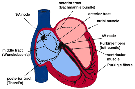
Figure 5. The conduction system of the heart. Normal excitation originates in the sinoatrial (SA)
node then propagates through both atria (internodal tracts shown as dashed lines). The atrial
depolarization spreads to the atrioventricular (AV) node, passes through the bundle of His (not
labeled), and then to the Purkinje fibers which make up the left and right bundle branches;
subsequently all ventricular muscle becomes activated.
|
|
|


