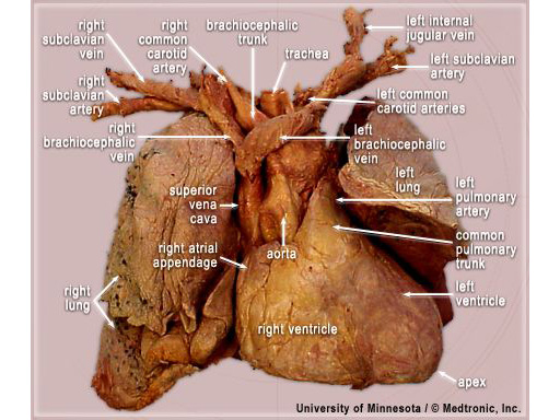
Anterior view of a "Heart-Lung Block" removed from a fixed cadaver. The
subclavian veins and arteries were kept intact connected to the great
vessels of the heart. This heart was enlarged and displaced the lower lobe
of the left lung.
Location:
the heart is positioned in the chest with 2/3 to the left of midline and
the inferior aspect is resting on the diaphragm. The apex of the heart is
pointing inferiorly and to the left. The media in this section display the
epicardial surface of the heart viewed either from the anterior, posterior
or left and right oblique aspects. Some of the media displays the heart in
a valentine position, with the long axis of the heart sitting in the
vertical plane. The rest of the images and videos display the heart
orientated in and attitudinally correct position, as the heart would be
viewed in the body, see anatomy
tutorial for more information.
Importance in cardiovascular diseases:
hypertrophic and dilated cardiomyopathies often alter the global structure
of the heart due to the heart changing size and shape during the progression
of the disease. Understanding how the heart is positioned in the body and
therefore where the relevant structures are is critically important for
cardiac surgery and bypass procedures.
Importance in device delivery:
epicardial pacing leads placed on both the atria and the ventricles will
often be used during trans-apical procedures and after bypass surgery to
control tachyarrhythmias and improve hemodynamic function in the presence
of arrhythmias.
Vista anterior de un "bloque corazón-pulmón" extraído de un cadáver fijo. Las venas y arterias subclavias se conservaron intactas conectadas a los grandes vasos del corazón. El corazón hipertrofiado desplaza el lóbulo inferior del pulmón izquierdo.
Localización:
el corazón se ubica en el tórax con 2/3 a la izquierda de la línea media y la pared inferior reposa sobre el diafragma. El vértice del corazón apunta hacia abajo y hacia la izquierda. Las imágenes de esta sección muestran la superficie epicárdica del corazón vista desde las caras anterior, posterior y oblicua izquierda y derecha. Algunas imágenes muestran el corazón en una posición de San Valentín, con el eje largo del corazón en el plano vertical. El resto de las imágenes y videos muestran el corazón en posición anatómica como se vería en el cuerpo; consulte el tutorial de anatomía para obtener más información.
Importancia en las enfermedades cardiovasculares:
Las miocardiopatías hipertróficas y dilatadas a menudo alteran la estructura global del corazón debido a que el corazón cambia de tamaño y forma durante la progresión de la enfermedad. Comprender cómo se posiciona el corazón en el cuerpo y, por lo tanto, dónde se encuentran las estructuras relevantes es de vital importancia durante la cirugía cardíaca y los procedimientos de derivación (bypass) cardiopulmonar.
Importancia en implante de dispositivos:
Los electrodos de estimulación epicárdica se pueden implantar tanto en las aurículas como en los ventrículos durante el abordaje transapical y después de la derivación (bypass) cardiopulmonar para controlar las taquiarritmias y mejorar la función hemodinámica en presencia de arritmias.