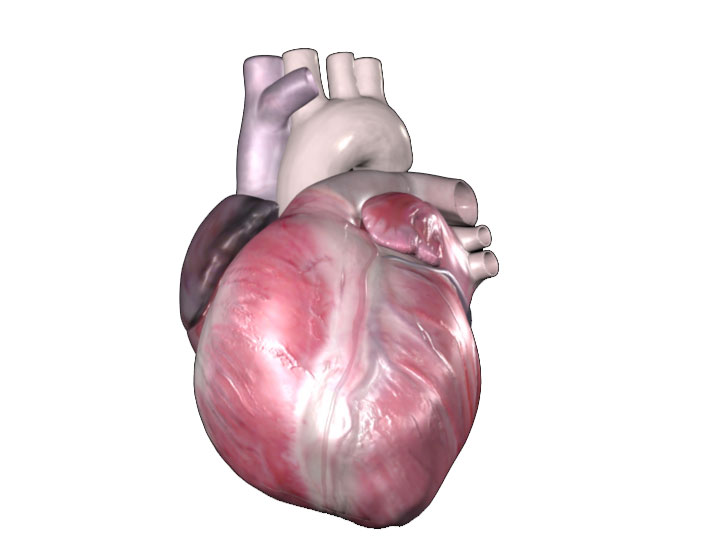
Importance in cardiovascular medicine:
The left aspect is a plane of the heart that becomes important during minimally
invasive trans-apical procedures due to the proximity of the heart’s apex
to the left wall of the chest cavity. The media display the heart in both the
valentine and attitudinally correct positions either in the body or explanted.
The pericardium, a double walled sac that protects and lubricates the heart,
can be seen in some of the media.
Important structures:
When positioned in the attitudinally correct orientation the left and right
sides of the heart can be seen with the left atrium and ventricle to the right
of the image or more posterior. The right atrium and ventricle can be seen to
the left of the image with the great vessels entering or exiting the heart to
the rear of the image. The apex of the heart can be seen at the front of the
image composed principally of the apex of the left ventricle.
Importancia en medicina cardiovascular:
El lado izquierdo es un plano del corazón que se vuelve importante durante los procedimientos transapicales mínimamente invasivos, debido a la proximidad del vértice del corazón con la pared izquierda del tórax. Las imágenes muestran el corazón tanto en la posición de San Valentín como en posición anatómica correcta, ya sea en el cuerpo o explantado. El pericardio es una membrana de doble pared que protege y lubrica el corazón, se puede apreciar en algunas imágenes.
Estructuras importantes:
Cuando se visualiza desde la posición anatómica correcta, se puede ver el lado izquierdo y derecho del corazón con la aurícula y el ventrículo izquierdo a la derecha de la imagen. La aurícula y el ventrículo derecho se pueden ver a la izquierda de la imagen y en la parte posterior se visualizan los grandes vasos que entran o salen del corazón. El vértice del corazón puede verse en la parte frontal de la imagen, compuesto principalmente por el vértice del ventrículo izquierdo.