Importance in cardiovascular medicine:
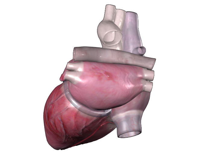
The posterior aspect is a plane in which the heart is not often viewed until the
heart is manipulated in the body during a surgical procedure. However, it can
be displayed in modern imaging modalities such as CT and MR and is used to help
orientate catheters and leads during catheter lab procedures. The media display
the heart in both the valentine and attitudinally correct positions either in
the body or explanted. The pericardium, a double walled sac that protects and
lubricates the heart, can be seen in some of the media.
Important structures:
When positioned in the attitudinally correct orientation the left atrium and
ventricle can be seen most prominently as they lie in a more posterior position
than the left. The left atrium can be seen to the right of the image with the
right superior and inferior pulmonary veins entering the chamber from the right
of the image and the left from the left. The coronary sinus can be seen
running around the base of the heart between the left atrium and ventricle and
the inferior vena cava can be seen to the bottom of the image connecting to the
inferior portion of the right atrium.
Importancia en medicina cardiovascular:
La pared posterior no se ve a menudo a menos que se manipule el corazón en el cuerpo durante un procedimiento quirúrgico. Sin embargo, se puede ver en imágenes como la TAC y RMN y se utiliza para ayudar a orientar los catéteres y los electrodos durante los procedimientos de hemodinamia. Las imágenes muestran el corazón tanto en la posición de San Valentín como en posición anatómica correcta, ya sea en el cuerpo o explantado. El pericardio es una membrana de doble pared que protege y lubrica el corazón, se puede apreciar en algunas imágenes.
Estructuras importantes:
Cuando se muestra el corazón en la posición anatómica, la aurícula y el ventrículo izquierdos se pueden ver de manera más prominente ya que se encuentran en una posición más posterior que las cavidades derechas. La aurícula izquierda se puede ver a la derecha de la imagen. Las venas pulmonares derechas se pueden ver ingresando a la aurícula desde la derecha de la imagen y las venas pulmonares izquierdas se pueden ver ingresando desde la izquierda de la imagen. Se puede apreciar el seno coronario que transcurre alrededor de la base del corazón entre la aurícula y el ventrículo izquierdos. La vena cava inferior se puede ver en la parte inferior de la imagen, se aprecia cómo conecta con la porción inferior de la aurícula derecha.