Importance in cardiovascular medicine:
The right aspect is the plane in which the heart is not often viewed until the
heart is manipulated in the body during a surgical procedure due to its
location near the core of the body. The media display the heart in both the
valentine and attitudinally correct positions either in the body or explanted.
The pericardium, a double walled sac that protects and lubricates the heart,
can be seen in some of the media.
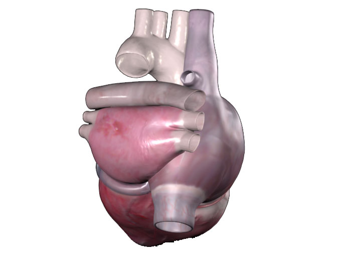
Important structures:
When positioned in the attitudinally correct orientation the left and right
atria are displayed most prominently. The left atria with the inferior and
superior left and right veins can be seen to the left of the image with the
pulmonary trunk running across the superior surface of the atrium. The arch
of the aorta can be seen to the top of the image and the superior and
inferior vena cava can be seen to the right of the image connecting to the
right atrium.
Importancia en medicina cardiovascular:
El lado derecho del corazón generalmente no se visualiza a menos que se manipule el corazón en el cuerpo durante un procedimiento quirúrgico. Las imágenes muestran el corazón tanto en la posición de San Valentín como en posición anatómica correcta, ya sea en el cuerpo o explantado. El pericardio es una membrana de doble pared que protege y lubrica el corazón, se puede apreciar en algunas imágenes.
Estructuras importantes:
Cuando se visualiza desde la posición anatómica correcta las aurículas izquierda y derecha son lo más prominente. A la izquierda de la imagen se puede visualizar la aurícula izquierda con las venas pulmonares inferiores y superiores y se observa el tronco pulmonar que transcurre en la superficie superior de la aurícula. El arco de la aorta se puede ver en la parte superior de la imagen. La vena cava superior e inferior se pueden ver a la derecha de la imagen conectando con la aurícula derecha.