University of Minnesota
http://www.umn.edu/
612-625-5000
Home
Make a Gift
VHLabs
 
HeartDatabase
 
Right Atrium
Right Ventricle
Pulmonary Trunk
Left Atrium
Left Ventricle
Aorta
Coronary Arteries
Cardiac Veins
External Images
MRI Images
Comparative Imaging
3D Modeling
Plastinates
 
Anatomy Tutorial
Cardiovascular Magnetic Resonance Tutorial
Comparative Anatomy Tutorial
Conduction System Tutorial
Congenital Defects Tutorial
Coronary System Tutorial
Device Tutorial
Echocardiography Tutorial
Physiology Tutorial
 
Project Methodologies
Cardiovascular Devices and Techniques at U of Minnesota
Acknowledgements
References and Links
Atlas in the media
 
Surgery Department
Principal


Coronary System Tutorial
What is the Coronary System? Coronary Arterial Anatomy Coronary Venous Anatomy Visualization of the Coronary System Biomedical Applications of the Coronary Arterial System Biomedical Applications of the Coronary Venous System Cardiac Venous Valves

What is the Coronary System

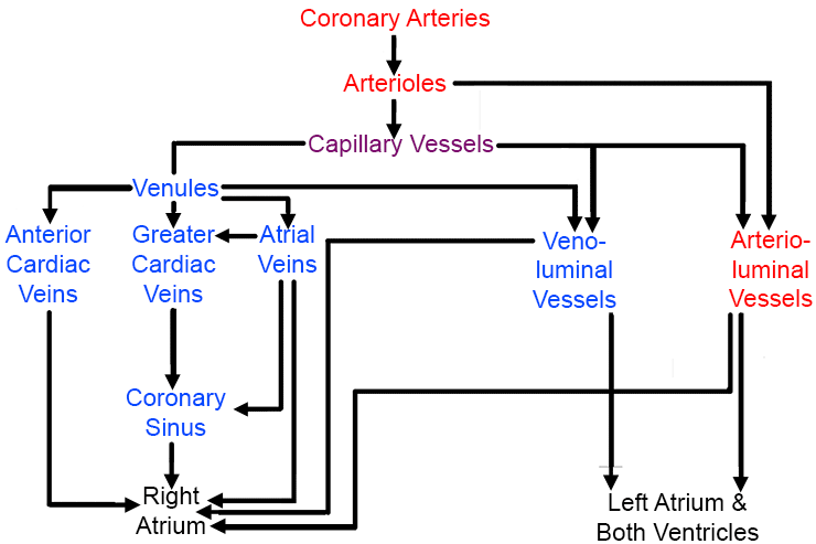
The coronary system is comprised of arteries, arterioles, capillaries, and cardiac veins and venules. The coronary arteries originate with right and left main coronary arteries which exit the ascending aorta just above the aortic valve. These two branches subdivide and course over the surface of the heart (epicardium) as they traverse away from the aorta. These arteries arborize into progressively smaller branches that progress inward to penetrate the epicardium and supply blood to the transmural myocardium. These coronary arteries branch into arterioles, which then branch into innumerable capillaries that deliver oxygenated blood to all of the heart's cells. Blood continues through the capillaries to begin the return back into the cardiac chambers via various drainage routes. The majority of capillaries drain into venules, which then empty into the coronary venous system. The coronary veins can be categorized into 2 subgroups: the greater and smaller cardiac venous system. The coronary veins return the deoxygenated blood back to the chambers of the heart.

Download movie: mp4

3-D reconstruction of contrast-computed tomography images for the coronary arterial and venous systems.

Supply and drainage diagram

The supply and drainage configuration of the coronary arterial and venous systems. [1]


References

  1. Spencer J, Anderson S, Iaizzo P (2013) Human Coronary Venous Anatomy for Interventions. J card trans res 6(2):208-217
 
 
© 2021 Regents of the University of Minnesota. All rights reserved. The University of Minnesota is an equal opportunity educator and employer. Privacy Statement