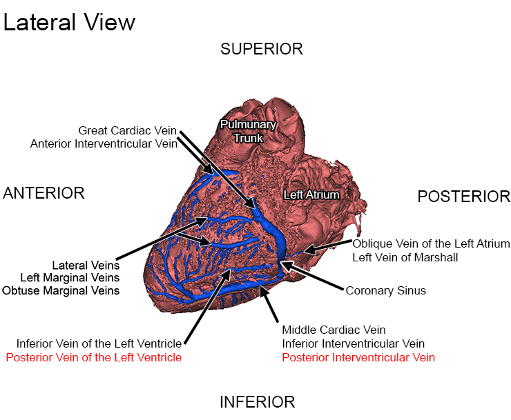
Anterior Interventricular Vein and Great Cardiac Vein
The anterior interventricular vein courses between the left and right ventricle on the anterior side of the heart. It typically begins around the apex and ends as it reaches the atrioventricular groove. The great cardiac vein, the longest venous vessel of the heart, consists of the anterior interventricular vein and its continuation along the atrioventricular groove [4,7]. As the great cardiac vein follows the left atrioventricular groove around the left side of the heart, the great cardiac vein is considered to be in close proximity to the anterolateral commissure of the mitral valve [9]. The great cardiac vein commonly continues until it merges with the coronary sinus [7].
Lateral Veins
The lateral veins, also known as the left marginal veins or the obtuse marginal veins, course along the left side of the heart and drain the left ventricular myocardium into the great cardiac vein or coronary sinus [7]. They are commonly located in an inferior position at an obtuse angle of the heart [10] and parallel the course of the left marginal branch of the left coronary artery [11].
Inferior Veins
The inferior veins of the left ventricle, previously known as the posterior veins of the left ventricle, typically originate from the lateral and inferior aspects of the left ventricle and course between the great cardiac vein and middle cardiac vein [4,7]. The vessels generally drain into the coronary sinus. Similar to the lateral veins, the anatomy of the inferior veins is also highly variable [4].
Middle Cardiac Vein
The middle cardiac vein, also referred to as the posterior interventricular vein or more correctly, the inferior interventricular vein, is a major coronary vein that typically originates near the apex and usually ascends in or very near to the posterior interventricular sulcus [4,6,7]. The middle cardiac vein drains into the coronary sinus or directly into the right atrium [4].
Small Cardiac Vein
The small cardiac vein, also known as the right cardiac vein [12], commonly drains the inferior and lateral wall of the right ventricle. A small vein in comparison to the previously mentioned veins, the small cardiac vein originates in the inferior part of the right coronary sulcus and courses the base of the right ventricle, paralleling the right coronary artery [7]. This vein typically empties into the coronary sinus, but sometimes drains into the middle cardiac vein or directly into the right atrium. The small cardiac vein is not always present in the human cardiac venous system [7].
Oblique Vein of the Left Atrium
The oblique vein of the left atrium, also referred to as Marshall's vein since it was first reported by John Marshall, delivers deoxygenated blood from the lateral and inferior regions of the left atrium to the atrioventricular groove [4]. As mentioned earlier, the termination of this vein is an anatomical landmark for the origin of the coronary sinus and the end of the great cardiac vein.
References
- Hutchins G, Moore G, Hatton E (1986) Arterial-venous relationships in the human left ventricular myocardium, Anatomic basis for countercurrent regulation of blood flow. Circulation 74:1195-1202
- Truex R, Angulo A (1952) Comparative study of the arterial and venous systems of the ventricular myocardium with special reference to the coronary sinus. Anat Rec 113:467-491
- Widmaider E, Raff H, Strang K (2004) Vander, Sherman, Luciano's human physiology, The mechanisms of body function (9 ed) McGraw-Hill, Boston
- Spencer J, Anderson S, Iaizzo P (2013) Human Coronary Venous Anatomy for Interventions. J card trans res 6(2):208-217
- Loukas, M et al (2009) Cardiac veins, a review of the literature Clinical Anatomy, 22(1):129-145
- Ho S, Sanchez-Quintana D, Becker A (2004) A review of the coronary venous system, a road less travelled. Heart Rhythm 1(1):107-112
- von Ludinghausen M (2003) The venous drainage of the human myocardium. Advances in anatomy, embryology, and cell biology 168:1-104
- Giudici M, Winston S, Kappler J et al (2002) Mapping the coronary sinus and great cardiac vein. Pacing Clin Electrophysiol 25:414-419
- El-Maasarany S, Ferrett C, Firth A, Sheppard M, Henin M (2005) The coronary sinus conduit function, anatomical study (relationship to adjacent structures). Europace 7:475-481
- Schaffler G, Groell R, Peichel K, Rienmuller R (2000) Imaging the coronary venous drainage system using electron-beam. CT Surg Radiol Anat 22:35-39
- Pejkovic B, Bogdanovic D (1992) The great cardiac vein. Surg Radiol Anat 14:23-28
- Williams P, Bannister L, Berry M (1995) Gray's anatomy. Churchill Livingston, London
|