|
Navigation within the coronary venous system has become increasingly important in commonly employed cardiovascular interventional procedures, including cardiac resynchronization therapy. While cannulating the coronary sinus ostium can be challenging due to the presence of the Thebesian valve, venous valves within the coronary venous system may hinder advancement of guide wires, catheters or pacing leads.(1) Venous valves within the major LV veins were prevalent in human hearts and were present most often at the ostia to smaller branch veins.(2,3) Anterior interventricular veins, posterior veins of the LV and posterior interventricular veins had more valves per vein than left marginal veins. While presence and development of these venous valves is variable, a venous valve in a patient could potentially complicate a cardiovascular interventional procedure.
- Karaca, M., Bilge, O., Hakan, D.M., et al. (2005). The anatomic barriers in the coronary sinus: Implications for clinical procedures. Journal of Interventional Cardiac Electrophysiology, 14(2):89-94.
- Anderson, S.E., Hill, A.J., Iaizzo, P.A. (2007). Venous valves: Unseen obstructions to coronary access. Journal of Interventional Cardiac Electrophysiology, 19:165-166.
- Anderson, S.E., Quill, J.L., Iaizzo, P.A. (2008). Venous valves within left ventricular coronary veins. Journal of Interventional Cardiac Electrophysiology, 23(2):95-9.
|
|
|
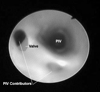
View from the base of the heart toward the apex along the length of the posterior interventricular vein (PIV). Two venous contributors can
be seen toward the bottom of the image. The ostia of the contributor to the upper left was partially covered by a venous valve.
|
|