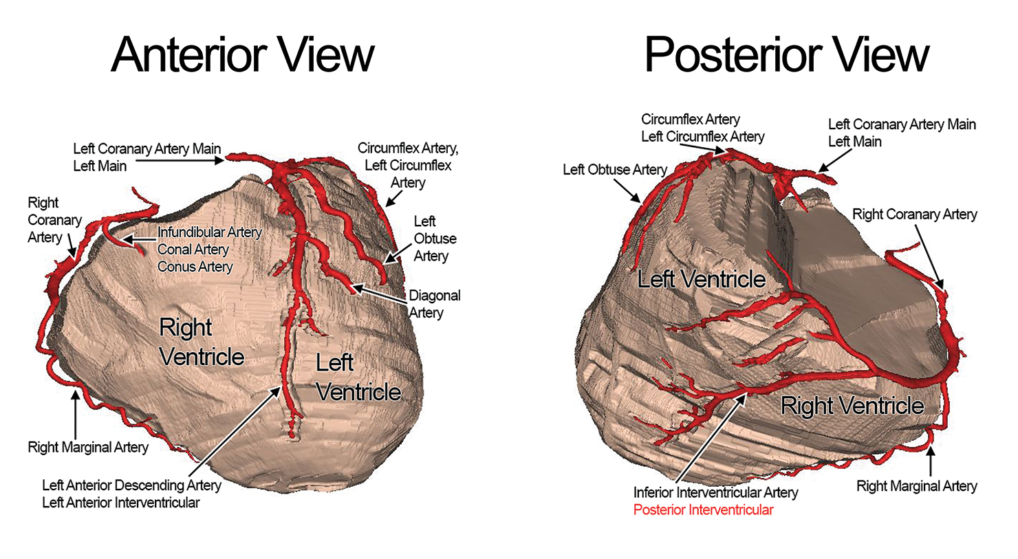
The Left Coronary Artery
The left coronary artery and its branches supply the majority of oxygenated blood to the ventricular myocardium and additionally to the left atrium, left atrial appendage, pulmonary artery, and aortic root [1, 2]. The left coronary artery main, also called the left main for short, typically originates at the left sinus of Valsalva and courses in the left anterior direction. After approximately 1 to 2 cm, the left main bifurcates into the left anterior descending artery and the circumflex artery between the left atrial appendage and the pulmonary trunk [3]. In some patients, there is a third artery that branches off the left main called the intermediate artery. This vessel usually supplies the left margin of the heart [3, 4].
The Left Anterior Descending Artery and its branches
The left anterior descending artery, also known as the anterior interventricular artery, originates at the end of the left main at an angle close to 180 degrees. The vessel curves around the pulmonary artery trunk and continues anteriorly down the interventricular septum to the apex of the heart. The left anterior descending artery typically branches into the diagonal arteries, deep septal perforators, and the left descending septal artery. Although there is a high amount of variation in the course of the diagonal arteries, they typically branch off at an angle and supply the free wall of the left ventricle. The deep septal perforators branch at 90 degree angles into the interventricular septum. These arteries typically supply the anterior two thirds of the septum and also have high variation. Finally, the left descending septal artery is an important vessel that supplies blood to the moderator band and anterior papillary muscle [3].
The Left Circumflex Artery and its branches
The circumflex artery, also referred to as the left circumflex artery, stems from the left main and courses along the left atrioventricular groove. The circumflex artery gives rise to small arterial branches that supply the aortic root and myocardium near the antrioventricular groove. In patients who are right dominant, the circumflex artery terminates into the left obtuse artery. The left obtuse artery courses down the left margin of the heart towards the apex. In patients who are left dominant (approximately one tenth of individuals), the circumflex continues along the atrioventricular groove until it terminates at the cardiac crux, where it branches into the artery that supplies the atrioventricular node [5, 6] and the inferior interventricular artery, traditionally known as the posterior interventricular artery [3].
The Right Coronary Artery and its branches
The right coronary artery originates in the right sinus of Valsalva and courses along the atrioventricular groove along the epicardial surface adjacent to the tricuspid valve annulus. The infundibular artery, also known as the conal artery, conus artery and third coronary artery, can also originate directly from the right sinus of Valsalva or as a branch of the right coronary artery. In three-fifth of the population, the next branch of the right coronary artery is the artery to the sinus node [3, 4]. This artery branches off the circumflex artery in the remaining two-fifths of the population [3, 4]. The right marginal artery is typically the largest branch of the right coronary artery. This major artery supplies the free wall of the right ventricle [3, 6]. In most individuals, the right coronary artery becomes the inferior interventricular artery when it reaches the diaphragmatic surface of the heart. Note that the right coronary artery does not typically taper in diameter until it gives rise to the inferior interventricular artery [7]. The inferior interventricular artery runs toward the apex of the left ventricle and supplies the diaphragmatic surface of the heart.
References
- von Ludinghausen, M (2003) The clinical anatomy of coronary arteries. Adv Anat Embryol Cell Biol 167(3-8):1-111
- Williams P, Bannister L, Berry M (1995) Gray's anatomy. Churchill Livingston, London
- Loukas M, Sharma A, Blaak C, Sorenson E, Mian A (2013) The Clinical Anatomy of the Coronary Arteries. J of Cardiovasc Trans Res 6:197-207
- Patel S (2008) Normal and anomalous anatomy of the coronary arteries. Semin Roentgenol 43:100-112
- Angelini P (1989) Normal and anomalous coronary arteries, Definitions and classification. Am Heart J 117:418-434
- James T (1961) Anatomy of the coronary arteries. Paul B Hoeber, New York
- Alexander R, Schlant R, Fuster V, O'Rourke R, Roberts R, Sonnenblick E (1999) Hurst's the heart. McGraw-Hill, New York
|