|
These models can be downloaded for you to print. If you need access to high-quality printed parts, these models can be printed through Stratasys Direct. We are proud to partner with Stratasys for our 3D printing needs as they have a strong commitment to supporting our education and outreach mission and can print in a wide variety of materials to fit any purpose.
|
|

|
|
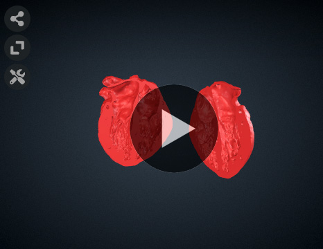
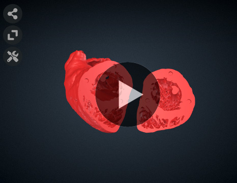
ME 4CH: This view offers a favorable acoustic window for visualizing the left atrium (LA), right atrium (RA), left ventricle (LV), right ventricle (RV), tricuspid valve, and mitral valve (A3A2-P2P1). It is an extensive view that facilitates the assessment of anatomy and function. It is particularly useful for evaluating primum atrial septal defects (ASD), ventricular septal defects (VSD), and abnormalities such as Ebstein anomaly.
|
|
|
|
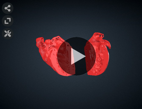
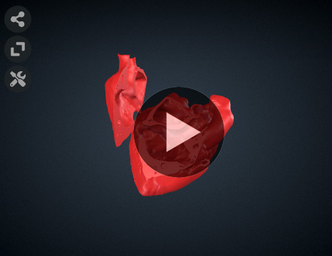
ME 2CH: The ME 2CH view is suitable for assessing regional left ventricular (LV) function, specifically the anterior and inferior walls, as well as the function of the mitral valve. It is one of the four recommended views for utilizing color Doppler across the mitral valve. The structures visible in this view include the left atrium (LA), left ventricle (LV), coronary sinus, mitral valve (P3-A3A2A1), and left atrial appendage.
|
|
|
|
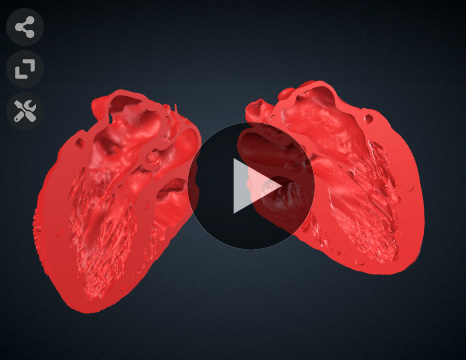
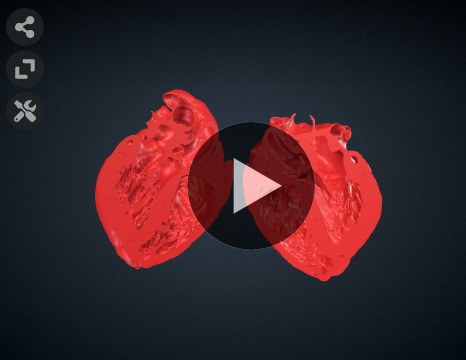
ME LAX: The ME LAX view allows visualization of the left atrium (LA), left ventricle (LV), left ventricular outflow tract (LVOT), right ventricular outflow tract (RVOT), aortic valve, mitral valve (P2-A2), and proximal ascending aorta. It is particularly useful for assessing regional left ventricular function in the inferolateral and anteroseptal walls, as well as the function of the mitral and aortic valves. Additionally, it provides appropriate visualization of membranous ventricular septal defects (VSD), and ascending aortopathy.
|
|
|
|
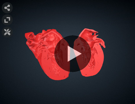
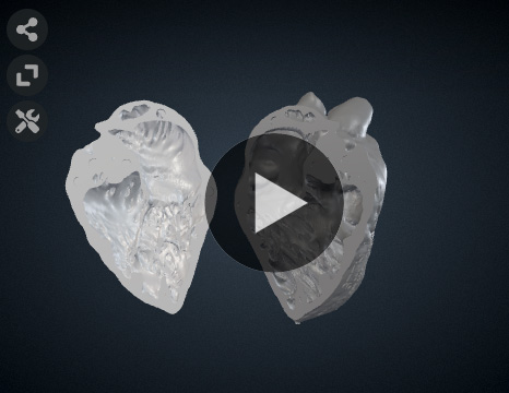
ME AV SAX: The ME AV SAX view provides a tri-leaflet view of the aortic valve, with the left, noncoronary, and right cusps visible in the posterior, right, and anterior parts of the ultrasound screen, respectively. This view also includes the right atrium (RA), left atrium (LA), superior interatrial septum, right ventricular outflow tract (RVOT), and pulmonary valves. It allows imaging of patent foramen ovale (PFO), atrial septal defects (ASD), as well as assessment of aortic and pulmonary valve function.
|
|
|
|
ME Mitral Comm.: The ME Mitral Comm. view enables focused analysis of the mitral valve structures, including P3-A3A2A1-P1, papillary muscles, and chordae tendineae. The left atrium (LA), left ventricle (LV), and coronary sinus are also visualized in this view. The diagnostic information obtained from this view includes assessment of mitral valve function and regional left ventricular function in the anterior/anterolateral and inferior/inferolateral walls.
|
|
|
|
TG Mid Papillary SAX view: The TG Mid Papillary SAX view offers diagnostic information regarding global left ventricular (LV) function, LV size, and volume status. It allows simultaneous visualization of the segments supplied by the left anterior descending (LAD), circumflex, and right coronary arteries, making it suitable for intraoperative monitoring of cardiac function. The structures visible in this view include the papillary muscles, left ventricle, and right ventricle.r and right ventricle.
|
|
|
|
ME bicaval view: The ME Bicaval view provides visualization of the left atrium (LA), right atrium (RA), right atrial appendage, interatrial septum, superior vena cava (SVC), and inferior vena cava (IVC). It offers valuable diagnostic information for detecting interatrial shunts, atrial septal defects (ASD), and confirming the position of intra-atrial catheters.
|
|
|
|
DeepTG 5CH view: The Deep TG 5CH view allows alignment of the Doppler beam with the aortic valve to observe transaortic flow and analyze peak velocities. It also permits interrogation of mitral valve function. In addition to the aortic and mitral valves, structures visible in this view include the left ventricle (LV), left ventricular outflow tract (LVOT), right ventricle (RV), and aortic valve.
|
|
|
|
Deep TG RV inflow view: The Deep TG RV Inflow view provides a clear visualization of the right ventricular (RV) anterior and inferior walls, papillary muscles, chordae tendineae, and tricuspid valve. With slight advancement, the proximal right ventricular outflow tract (RVOT) becomes visible, allowing Doppler imaging through it.
|
|
|
Bibliography:
Hahn RT, Abraham T, Adams MS, et al. Guidelines for performing a comprehensive transesophageal echocardiographic examination: Recommendations from the American Society of Echocardiography and the Society of Cardiovascular Anesthesiologists. Journal of the American Society of Echocardiography. 2013;26(9):921-964. doi:10.1016/j.echo.2013.07.009
|