|
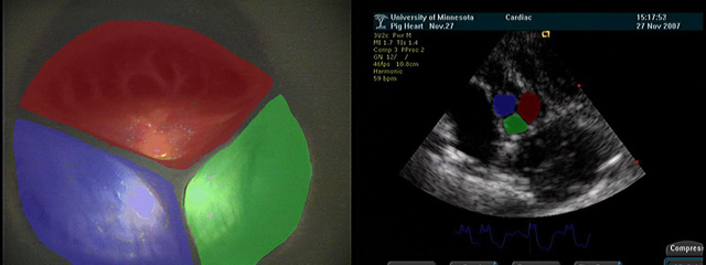
The aortic valve controls the flow of blood leaving the left ventricle
and has three cusps: the right coronary cusp, the left coronary cusp,
and the non-coronary cusp. The top, left image shows the short axis view
of the aortic valve under direct visualization, with the cusps highlighted
in red, green, and blue, respectively. The bottom, left video shows the
direct visualization of the aortic valve, as viewed from the aorta,
throughout the cardiac cycle. The corresponding echocardiography view
is shown in the upper right image, with the valve cusps colored using the
same scheme. Finally, the bottom, right video shows a short axis view of
the aortic valve using transthoracic echocardiography.
The long axis view of the aortic valve allows for imaging of several
important anatomical features. These include the aortic valve leaflets,
the sinus of Valsalva, and the sinotubular junction. Color flow
echocardiography can be utilized to visualize the flow through the aortic
valve in this view.
The sinus of Valsalva contains two ostia; the left coronary ostium and
the right coronary ostium. The left coronary ostium immediately branches
into the left anterior descending coronary artery and the circumflex
artery. Both of these ostia can be viewed using an appropriately angled
short axis echocardiography view.

|


