|
The right and left ventricles of all large mammals are structurally similar and
contain the same components (1). The walls of the ventricles are significantly
more muscular than those of the atria and the walls of the left ventricle are
significantly thicker than those of the right. Near the apex, the inner walls of
the ventricles have muscular ridges called trabeculae carnae; the extent of
trabeculation varies between humans and animals (3, 9, 10, 11). The ventricular
walls also have papillary muscles that support the atrioventricular valves
through the chordae tendineae. The number of these muscles can vary both within
and between species (3). Both ventricles may also contain cross-chamber fibrous
or muscular bands that typically carry Purkinje fibers. The moderator band is a
prominent band within the right ventricle that is common to humans, dogs, pigs
and sheep though its origin and insertion points, as well as its structure, can
vary between species (3).
Human
The trabeculations of the right and left ventricles in the human heart are
both more numerous and finer than those seen in canine, ovine or porcine hearts
(3, 8). The moderator band of the right ventricle is generally a free arc in
the human heart but can also be in the form of a ridge (1). In humans, the
moderator band arises more apically on the septal wall than seen in canine,
porcine, or ovine hearts and inserts at the base of the anterior papillary
muscle. Left ventricular bands are also typically present in the human heart
(1). Within the right ventricle, human hearts have one well defined anterior
papillary muscle, one to three posterior papillary muscles and one to three
septal papillary muscles (1). In the left ventricle, human hearts have one to
three anterior papillary muscles and one or two posterior papillary muscles. The
right ventricle inflow tract in humans is significantly larger than that of the
canine, ovine or porcine heart (1). The right ventricular outflow tract however
is similar in size to that of the canine, ovine and porcine heart. The left
ventricular inflow tract of humans is similar in length to ovine and porcine
hearts but is significantly longer than in canine hearts (1). The left
ventricular outflow tract of the human heart however is generally larger than in
canine, ovine or porcine hearts (1).
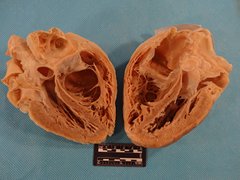
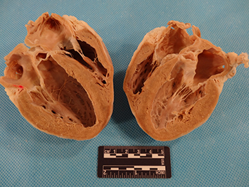
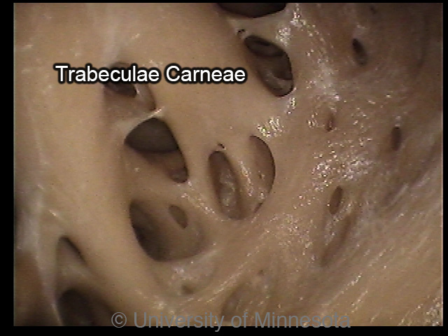
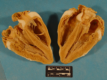
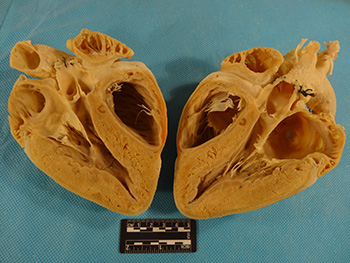
A human heart cut coronally to form an anterosuperior half (left) and a posteroinferior half (right). The trabeculae carneae in the apex can be seen here. Notably, they are both more numerous and finer than in the hearts of swine, canines or sheep (3, 8).
|
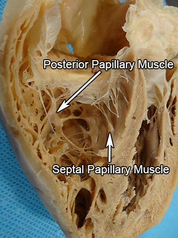
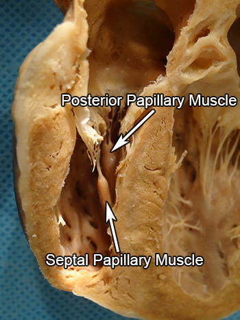
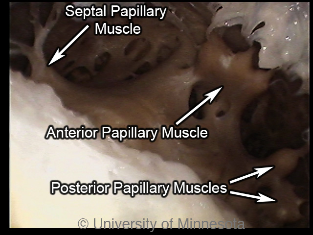
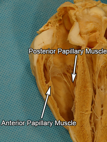
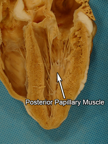
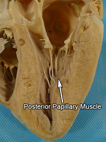
The posterioinferior side of a plastinated human right ventricle is pictured here. Human hearts may have one to three posterior papillary muscles and one to three septal papillary muscles (1). This particular heart has one posterior papillary muscle. One of the heart’s two septal papillary muscles is visible here.
|
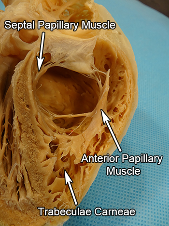
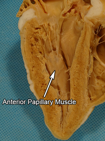
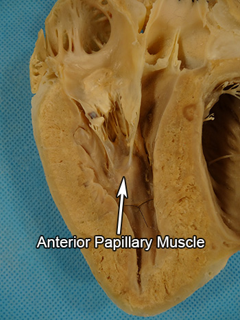
The anterosuperior side of a plastinated human right ventricle is pictured here. The human heart has one well defined anterior papillary muscle and one to three septal papillary muscles (1). The heart’s anterior papillary muscle is visible and one of this heart’s two septal papillary muscles can be seen here.
|
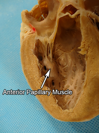
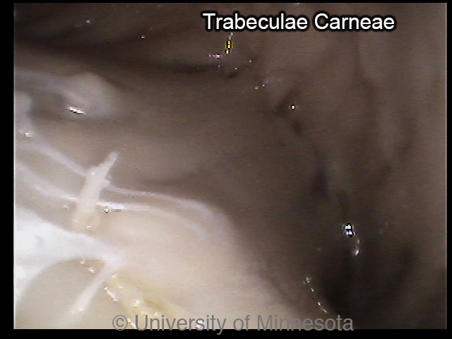
An internal image of a human right ventricle. Human hearts have one well defined anterior papillary muscle on the free wall of the ventricle (1). The numerous trabeculae of the human heart are also visible (3, 8).
|
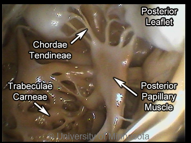
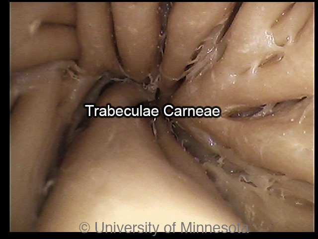
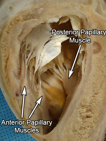
An internal image of the human heart right ventricle. Humans may have one to three posterior papillary muscles within the right ventricle (1). This heart’s only posterior papillary muscle and trabeculae carneae can be seen here as well as the chordae tendineae that connect the papillary muscle and the tricuspid valve.
|
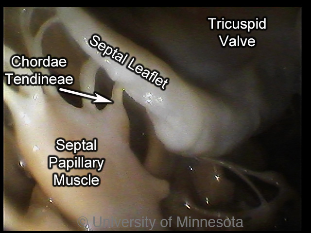
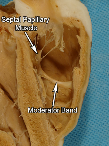
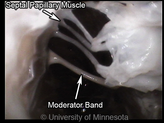
An internal image of the human right ventricle. Humans may have one to three septal papillary muscles (1). The septal papillary muscle and the chordae tendineae connecting it to the tricuspid valve’s septal leaflet can be seen.
|
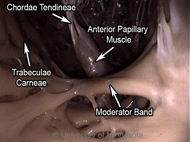
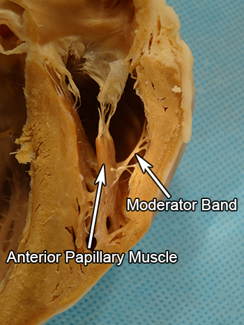
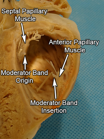
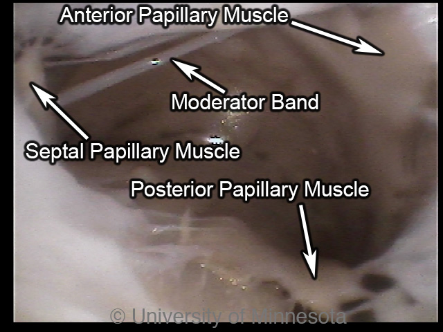
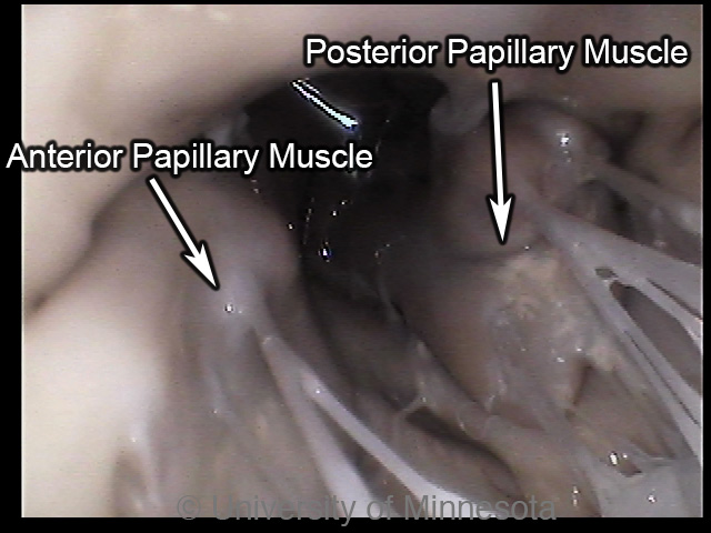
An internal image of the human right ventricle. Visible are the anterior papillary muscle, its chardae tendineae connecting it to the tricuspid valve, the trabeculae carneae and the moderator band. The moderator band of humans arises apically on the septal wall and originates at the base of the anterior papillary muscle (1).
|
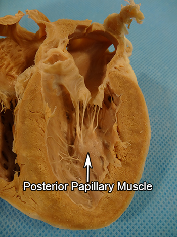
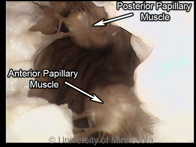
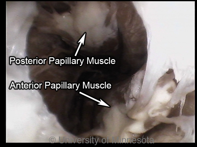
The posterior side of a plastinated human left atrium is pictured here. Humans have a single anterior papillary muscle and a single posterior papillary muscle in the left ventricle (1). Both the posterior and anterior papillary muscles are visible here.
|
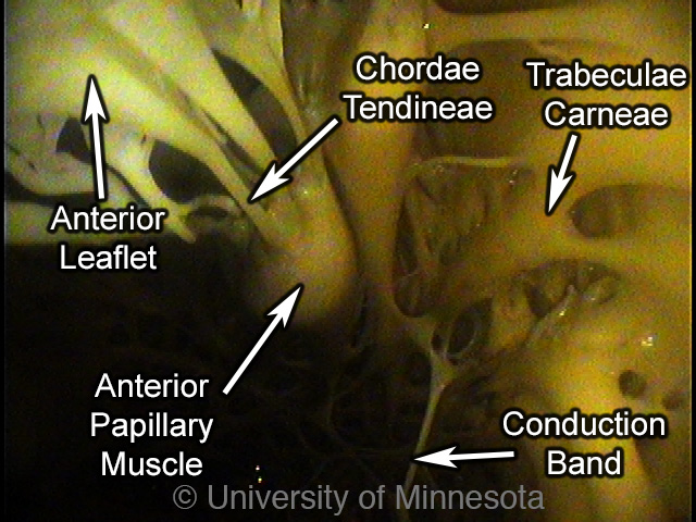
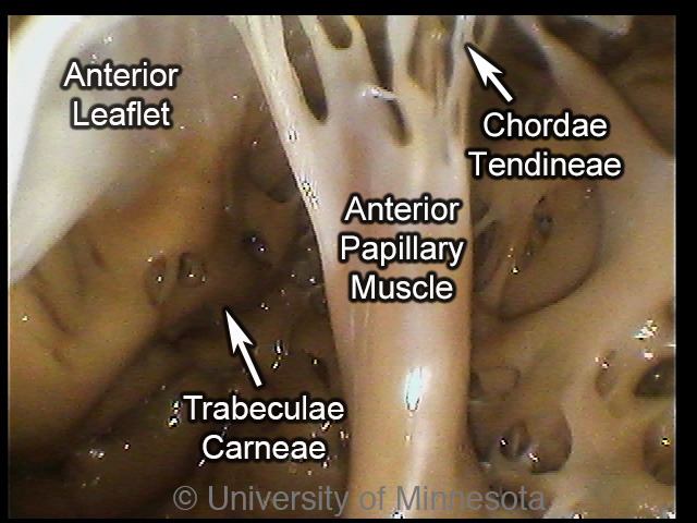
A view of the inside of a human left ventricle from above. Visible are the anterior papillary muscle, supporting the anterior leaflet of the mitral valve, the trabeculae carneae and a conduction band.
|
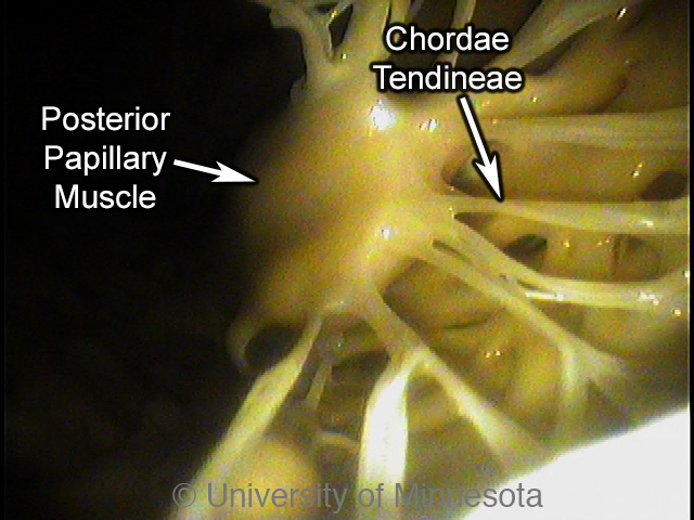
A internal image of the human left ventricle taken from a superior position. The posterior papillary muscle can be seen as well as the chordae tendineae connecting it to the mitral valve.
|
Canine
The trabeculae carneae of the right and left ventricles in the canine
heart are both fewer and coarser compared to those seen in the human heart (1).
Notably, the right ventricle of the canine heart has more trabeculation compared
to porcine and ovine hearts though (1). However, the left ventricles of these
three animals have similar trabeculation (1). A moderator band is present in the
right ventricle as a free arching fibrous network that originates at the
anterior papillary muscle or septal wall and inserts on the free walls of the
right ventricle at multiple sites (1). Within the right ventricle, canine hearts
can have one or two anterior papillary muscles, one to three posterior papillary
muscles and one to three or more septal papillary muscles (1). In the left
ventricle, the canine heart has a single anterior papillary muscle and a single
posterior papillary muscle. The right ventricle inflow tract is significantly
shorter than in humans but is similar in length to ovine and porcine hearts (1).
The right ventricular outflow tract is similar in length to those of human,
porcine and ovine hearts. The left ventricle inflow tract of the canine heart is
significantly shorter than that of the human heart, but similar in length to
swine and sheep (1). Likewise, the left ventricular outflow tract is
significantly smaller than in humans but similar in length to swine and sheep.
A plastinated canine heart cut coronally with the anterior half on the left and the posterior half on the right. The canine heart trabeculae carneae can be seen which are coarser than those of humans. Compared to swine and sheep hearts, the right ventricle has greater trabeculation though the left has similar trabeculation compared to these other animals (1).
|
The posterior half of a plastinated canine right ventricle is pictured here. Canine hearts have one or two anterior papillary muscles, one to three posterior papillary muscles and one to three septal papillary muscles supporting the tricuspid valve (1). This particular heart contains one of each papillary muscle. The septal and posterior papillary muscles are visible here.
|
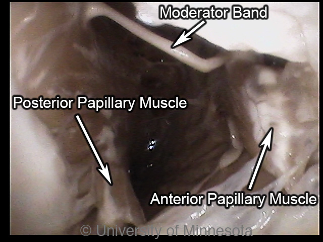
The anterior half of a plastinated canine right ventricle can be seen here. Canine hearts have one or two anterior papillary muscles, one to three posterior papillary muscles and one to three septal papillary muscles supporting the tricuspid valve (1). This particular heart contains one of each papillary muscle. The anterior papillary muscle is visible here as well as the moderator band, originating on the anterior papillary muscle and inserting at multiple points on the free wall.
|
A internal picture of a canine right ventricle taken with the camera in the right atrium looking inferiorly. Canine hearts have one or two anterior papillary muscles, one to three posterior papillary muscles and one to three septal papillary muscles (1). This particular heart has a single septal papillary muscle, one anterior papillary muscle and two posterior papillary muscles.
|
The trabeculae carneae of a canine heart are pictured here. The trabeculae of canines are fewer and courser compared to humans but are more abundant in the right ventricle compared to ovine and swine hearts (1).
|
The posterior half of a plastinated canine left ventricle is pictured here. There is one anterior and one posterior papillary muscle in the left ventricle of canines (1). The posterior papillary muscle, supporting the mitral valve, and the trabeculae carneae are also visible. Compared to humans, the trabeculae of canines are fewer and courser (1).
|
The anterior half of a plastinated canine left ventricle is pictured here. There is one anterior and one posterior papillary muscle in the left ventricle of canines (1).The anterior papillary muscle supporting the mitral valve is visible as well as the trabeculae. Compared to humans, the trabeculae of canines are fewer and courser (1).
|
A internal view of a canine left ventricle with the camera positioned in the left atrium looking inferiorly. There is one anterior and one posterior papillary muscle in the left ventricle of canines (1). Both papillary muscles are visible here as well as the trabeculae carneae which are fewer and coarser compared to humans (1)
|
A closer look at the left ventricular apex of a canine heart. The trabeculeae of dogs, like sheep and pigs, are courser compared to those in human hearts. Also visible are fibrous bands of the left ventricle.
|
|
Ovine
The trabeculae carneae of the right and left ventricles in the ovine heart
are fewer and coarser compared to those seen in the human heart (3, 8).
Moderator bands are present in the right ventricle as a free arching muscular
structure that originates on or near the septal papillary muscle and inserts at
the body or head of the anterior papillary muscle (1). In the right ventricle,
the ovine heart has a single anterior papillary muscle, one or occasionally two
posterior papillary muscle and one or two septal papillary muscles (1). In the
left ventricle, ovine hearts have a single anterior papillary muscle and one or
occasionally two posterior papillary muscles. The right ventricle inflow tract
is significantly shorter compared to humans but is similar in size to canine and
porcine hearts (1). The right ventricle outflow tract is comparable in length to
human, porcine and canine hearts. The left ventricle inflow tract is similar in
length to human, canine and porcine hearts (1). The left ventricle outflow tract
of the ovine heart is significantly shorter than the human heart but is similar
to the other animals' hearts.
A plastinated ovine heart cut coronally, forming a anterior half (left) and a posterior half (right). The papillary muscles, trabeculae carneae and overall structure of the right and left ventricles can be seen. The trabeculae carneae are noticeably fewer and courser compared to those in human hearts.
|
The posterior half of the right ventricle in a plastinated ovine heart. Ovine hearts have one anterior papillary muscle, one or two posterior papillary muscles and one or two septal papillary muscles (1).This heart’s one posterior and one anterior muscles are visible, supporting the tricuspid valve through the chordate tendineae. The trabeculae carneae are noticeably fewer and courser compared to human hearts (3, 8).
|
The anterior half of the right ventricle of a plastinated ovine heart. Ovine hearts have one anterior papillary muscle, one or two posterior papillary muscles and one or two septal papillary muscles (1). This heart’s septal papillary muscle is visible, supporting the tricuspid valve through its chordae tendineae. The moderator band, originating at the base of the septal papillary muscle and inserting at the base of the anterior papillary muscle, is also visible.
|
A internal image of a ovine right ventricle, viewed superiorly. Ovine hearts have one anterior papillary muscle, one or two posterior papillary muscles and one or two septal papillary muscles (1). The heart’s anterior papillary muscle and its one posterior papillary muscle are visible connecting to the tricuspid valve via the chordae tendineae. The moderator band is also visible, originating at the base of the septal papillary muscle and inserting near the anterior papillary muscle.
|
A closer look at the apex of a ovine right ventricle. The trabeculae carneae are visible and are noticeably fewer and courser compared to the hearts of humans.
|
A superior view of the septal papillary muscle and moderator band in a ovine heart. The moderator band originates in the septal wall by the septal papillary muscle, crosses the right ventricle as a free arc, and inserts near the anterior papillary muscle.
|
The posterior half of the left ventricle of a plastinated ovine heart. Ovine hearts have a single anterior papillary muscle and may have one or two posterior papillary muscles in the left ventricle (1). This particular heart has one posterior papillary muscle, visible here, supporting the mitral valve through its chordae tendineae.
|
The anterior half of the left atrium of a plastinated ovine heart. Ovine hearts have one anterior papillary muscle and may have one or two posterior papillary muscles (1). The anterior papillary muscle and its chordate tendineae supporting the mitral valve can be seen.
|
A internal view of the ovine left ventricle. The camera is positioned in the left atrium and is looking inferiorly into the ventricle. The ovine heart has one anterior papillary muscle and one or two posterior papillary muscles supporting the mitral valve (1). This particular heart has one anterior papillary muscle and one papillary muscle.
|
A view of the apex within the left ventricle of a ovine heart. The hearts of sheep, like the other animals here, have much coarser trabeculae carneae than the hearts of humans. Also visible are the conduction bands that help propagate the signal for contraction through the left ventricle.
|
Porcine
The trabeculae carneae of the right and left ventricles in the porcine
heart are both fewer and coarser compared to those seen in the human heart (3,
8). The moderator band is present in the right ventricle as a free arching
structure that originates on or near the septal papillary muscle and inserts
into the body or head of the anterior papillary muscle (1). In the right
ventricle, the porcine heart has a single anterior papillary muscle, one to
three posterior papillary muscles and one to three septal papillary muscles (1).
In the left ventricle, the porcine heart has a single anterior papillary muscle
and a single posterior papillary muscle. The right ventricle inflow tract in the
porcine heart is significantly shorter than the human heart but is similar in
size to canine and ovine hearts (1). The right ventricle outflow tract however
is similar in length to the human heart as well as to canine and ovine hearts.
The left ventricle inflow tract is similar in length to human, canine and ovine
hearts (1). The left ventricle outflow tract is shorter than in the human heart
but similar in length compared to canine and ovine hearts (1).
A plastinated porcine heart cut coronally into a anterior half (left) and a posterior half (right). The trabeculae carneae, papillary muscles and overall structure of the right and left ventricle can be seen. The trabeculae carneae of swine hearts are noticeably fewer and coarser compared to humans (1).
|
The posterior half of the right ventricle of a plastinated porcine heart. Swine have a single anterior papillary muscle, one to three posterior papillary muscles and one to three septal papillary muscles in the right ventricle (1). This particular heart has one posterior papillary muscle and one anterior papillary muscle visible here.
|
The anterior half of the right ventricle of a plastinated porcine heart. Swine have a single anterior papillary muscle, one to three posterior papillary muscles and one to three septal papillary muscles in the right ventricle (1). This particular heart has one septal papillary muscle and part of its one anterior papillary muscle visible here. Though the moderator band has been broken in this heart, the origin on the septal papillary muscle and the insertion on the anterior papillary muscle are visible.
|
A internal view of a porcine right ventricle. The camera is positioned in the right atrium and is looking inferiorly into the ventricle. The right ventricle of the swine heart has one anterior papillary muscle, one to three posterior papillary muscles and one to three septal papillary muscles (1). This particular heart has one anterior, one posterior and one septal papillary muscle. Also visible is the moderator band which is present in swine hearts as a free arching structure that originates on or near the septal papillary muscle and inserts on the anterior papillary muscle (1).
|
A internal view of the apex of a porcine right ventricle. The trabeculae carneae of the swine heart, like the hearts of dogs and sheep, are coarser than in the hearts of humans (1).
|
The posterior half of the left ventricle of a plastinated porcine heart. Swine have one anterior papillary muscle and one posterior papillary muscle supporting the mitral valve (1). The posterior papillary muscle is visible here.
|
The anterior half of the left ventricle of a plastinated porcine heart. Porcine hearts have one anterior papillary muscle and one posterior papillary muscle supporting the mitral valve (1). The anterior papillary muscle is visible here.
|
A internal view of the left ventricle of a porcine heart. The camera is positioned just inferior to the mitral valve and looking inferiorly. Swine hearts have one anterior papillary muscle and one posterior papillary muscle in their left ventricle both of which are visible here (1).
|
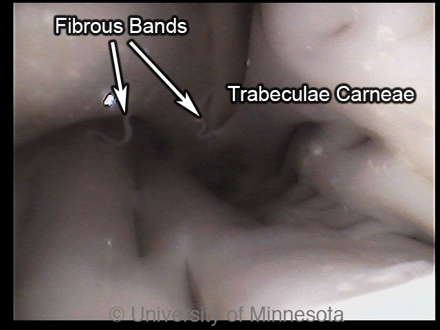
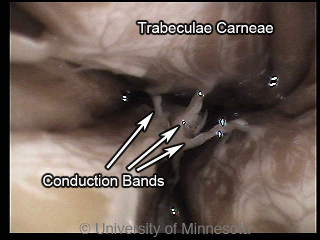
An internal image of the apex of a porcine heart. The trabeculae carneae in pigs, similar to the hearts of swine and canine, are much coarser than in human hearts (1). Also visible are two fibrous bands within the left ventricle.
|
|
|


