|
The hearts of large mammals have four cardiac valves with similar structures and
locations. Left and right atrioventricular valves sit between the atria and
ventricle on both sides of the heart while two semilunar valves separate the
right and left ventricles from the pulmonary artery and aorta respectively.
While the structures of each valve are largely similar, variations in the valves
exist that distinguish the valves of different species.
(terms in parenthesis describe attitudinally correct terms for describing the
human heart.)
Tricuspid Valve
Named for its three major cusps, this valve separates the right atrium from the
right ventricle. Papillary muscles attach to the cusps via chordae tendinae and
prevent the prolapsed of leaflets into the atria. Interspecies and
interindividual differences exist in the total number of papillary muscles
supporting the valve. Canine tricuspid valves are noticeably different from
those of humans, swine and sheep in that the commissures between the
anteriosuperior leaflet and the inferior leaflets can be fused, giving the
appearance of having only two cusps (9). While both the anterior (superior) to
posterior (inferior) diameter and the septal (posterior) to lateral (anterior)
diameter are largely similar between humans and these model animals, the
anterior to posterior diameter is noticeably larger in humans compared to swine.
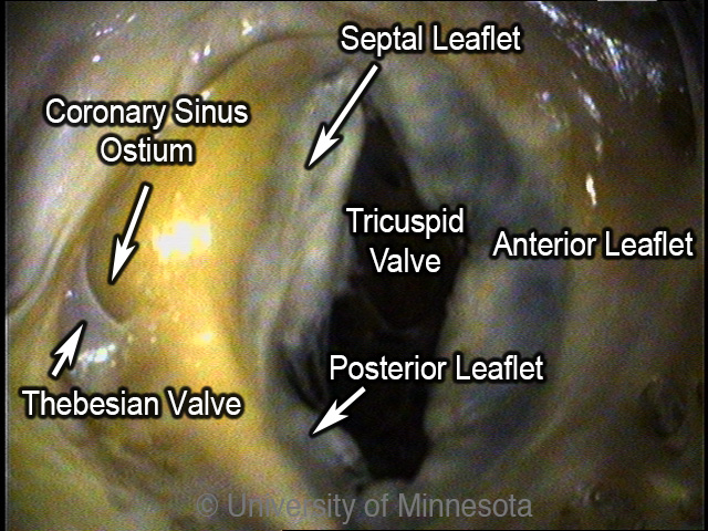
A human tricuspid valve viewed from the right atrium. The anterior (superior) to posterior (inferior) diameter is larger in humans than in swine but is similar in length to canine and ovine tricuspid valves (1). The septral (posterior) to lateral (anterior diameter is similar to the animal hearts here (1).
|
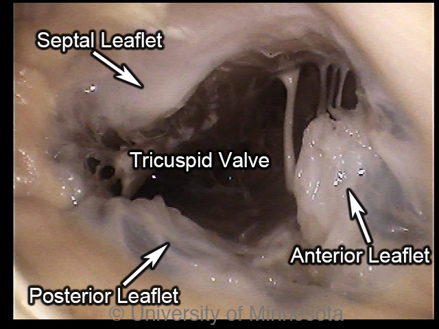
A canine tricuspid valve viewed from the right atrium. The anterior (superior) to posterior (inferior) diameter and the septal (posterior) to lateral (anterior) diameter is similar in length in dogs to the diameters in human, swine and ovine tricuspid valves (1). Canine tricuspid valves are notably in that the commissure between the anterosuperior leaflet and the inferior leaflets may be fused, giving the appearance of only two leaflets (9).
|
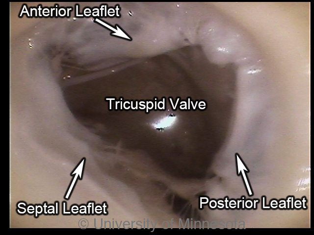
A ovine tricuspid valve viewed from the right atrium. The anterior (superior) to posterior (inferior) diameter and the septal (posterior) to lateral (anterior) diameter of the ovine tricuspid valve is similar in length to the diameter in human, swine and canine tricuspid valves (1).
|
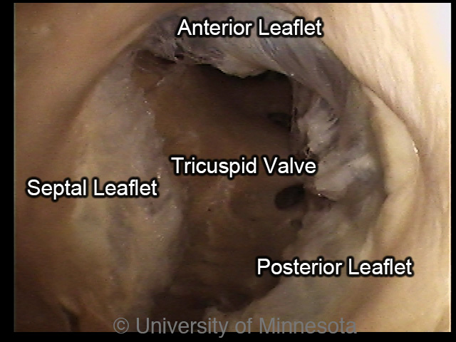
A swine tricuspid valve viewed from the right atrium. The anterior (superior) to posterior (inferior) diameter is smaller in swine than in humans but is comparable to the diameter in sheep and dogs. The septal (posterior) to lateral (anterior) diameter is similar in length in pigs to the diameter in human, ovine and canine tricuspid valves.
|
Mitral (Bicuspid) Valve
Named for its two major cusps, this valve separates the left atrium from the
left ventricle. Aside from the two major cusps that are always present, a
variable number of small commissural cusps, also called scallops, exist in large
mammals giving the impression of extra leaflets (3). Differences also exist
between species in the composition of the leaflets and in the fibrous ring that
supports the mitral valve. For example, a segment of the fibrous ring is visible
in humans at the base of the mural cusp however; this structure is difficult to
distinguish in certain breeds of dogs and is inconspicuous in sheep (25).
Regarding the size of the Mitral valve, the anterior (superior) to posterior
(inferior) diameter of the mitral valve is similar in length between humans,
canines, swine and sheep. The septal (posterior) to lateral (anterior) diameter
is significantly longer in humans compared to the animal models, which have
similar measurements.

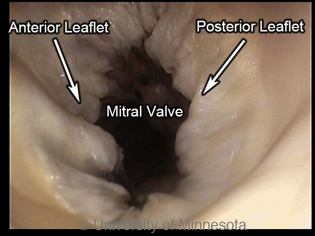
A human mitral valve viewed from the left atrium. In humans, a segment of the supporting fibrous ring is visible at the base of the mural cusp (25). The anterior (superior) to posterior (inferior) distance of the mitral valve is similar in length in human, swine, canine, and ovine hearts but the septal (posterior) to lateral (anterior) diameter is longer in humans compared to the animal hearts here (1).
|
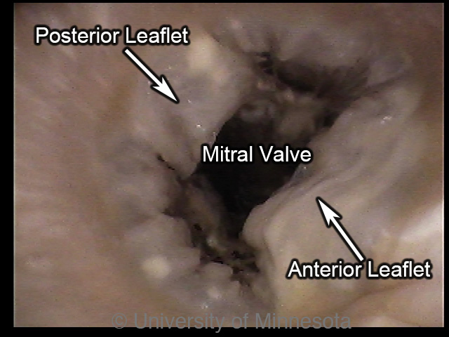
A canine mitral valve viewed from the left atrium. In certain breeds of dogs, the supporting fibrous ring below the mural cusp is difficult to distinguish (25). The anterior (superior) to posterior (inferior) distance of the mitral valve is similar in canine, human, swine and ovine hearts (1). The septal (posterior) to lateral (anterior) diameter shorter than in humans but is similar in length to swine and ovine mitral valves (1).
|
A ovine mitral valve viewed from the left atrium. The supporting fibrous ring below the mural cusp is difficult to distinguish in the hearts of sheep (25). The anterior (superior) to posterior (inferior) distance of the mitral valve is similar in ovine, human, swine and canine hearts (1). The septal (posterior) to lateral (anterior) diameter is shorter than in humans but is similar in length to swine and canine mitral valves (1).
|
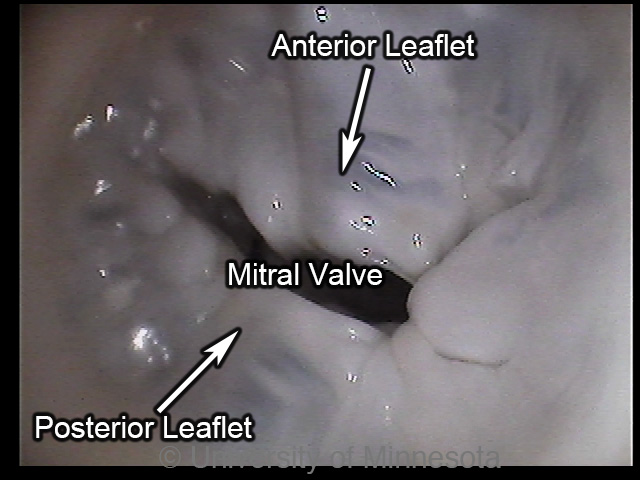
A swine mitral valve viewed from the left atrium. The anterior (superior) to posterior (inferior) distance of the mitral valve is similar in swine, human, sheep and canine hearts (1). The septal (posterior) to lateral (anterior) diameter is shorter than in humans but is similar in length to sheep and canine mitral valves (1).
|
Aortic Valve
The aortic valve separates the left ventricle from the aorta, preventing the
backflow of blood into the heart during ventricular relaxation. The valve is
formed by three cusps, each one evenly spaced around the valve's
circumference. Differences in leaflet shape and structure exist between species.
For example, the noncoronary cusp of swine is noticeably smaller than its other
two, while the three cusps of human, canine and sheep aortic valves are largely
uniform in size (26). Another significant anatomical difference is in the aortic
valves of sheep where the cusps are substantially thinner compared to those of
humans, swine or canine (26). Differences also exist in the structural support
around the valve. Notably, humans have much less muscular attachment surrounding
the valve compared to animal hearts (26). Human aortic valves also tend be have
a wider diameter compared to the animal hearts here and the canine aortic valve
is notably smaller in diameter than swine or sheep, which are similar in size (1).
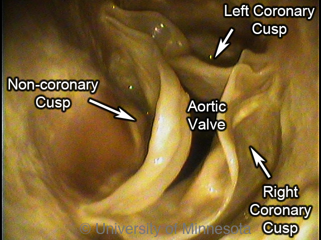
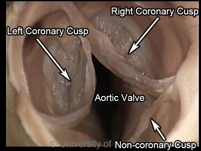
A human aortic valve viewed with the camera positioned in the ascending aorta. The human aortic valve has less muscular support than in swine, canine or sheep aortic valves but has a larger diameter compared to these animal valves (26, 1).
|
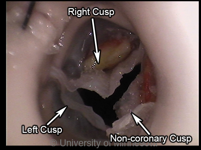
A canine aortic valve viewed from the aortic root. The canine aortic valve, like the other animal hearts here, has more muscular support compared to the aortic valves of humans (26). The diameter of the canine aortic valve is also smaller than the aortic valves of humans, swine or sheep (1).
|
A ovine aortic valve viewed from the aortic root. The aortic valve of sheep, like the other animal hearts here, has greater muscular support than the human aortic valve (26). The diameter of the sheep aortic valve is smaller than in humans, larger than in canine and similar in size to swine (1). Notably, the cusps of the sheep aortic valve are substantially thinner than in the other hearts here (26).
|
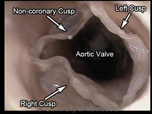
A porcine aortic valve viewed from the aortic root. The aortic valve of swine, like the other animal hearts here, has greater muscular support than the human aortic valve (26). The diameter of the swine aortic valve is smaller than in humans, larger than in canine and similar in size to sheep (1). Notably, the non-coronary cusp is smaller than the right and left cusps, unlike in humans, sheep or canine where all three cusps are similar in size (26).
|
Intervalvar septum (membranous septum)
The intervalvar septum is a fibrous continuity between the mitral and aortic
valves that extends from the central fibrous body to the left fibrous trigone
(3). The length of the septum varies between species but is absent in sheep (25).
|


