|
The right and left atria are located at the base of the heart and are separated
from each other by the interatrial septum and from the ventricles by the cardiac
skeleton (7). Structures common to all large mammalian atria include the sinus
venosus, the crista terminalis, the fossa ovalis, the Eustachian valve, the
Thebesian valve and right and left atrial appendages (3, 8, 9, 10, 11). However,
these structures can vary between species in their relative locations within the
atria or in their overall shape and size (3, 8). All large mammalian hearts also
have two vena cava returning blood from the body to the right atrium whose ostia
vary in relative locations between humans and animals (3, 8, 10). Pulmonary
veins, the number of which varies between individuals as well as species, return
blood to the left atrium from the pulmonary circulation in mammalian hearts (8,
9, 12).
(terms in parenthesis describe attitudinally correct terms for describing the
human heart.)
Human
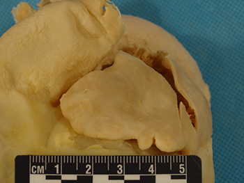
In the human heart, the free portion of the right atrial appendage is
usually triangular in shape while the left is generally tubular (1). Either the
right or the left atrial appendage may be larger than the other. The vena cava
of the human heart enter the right atrium in-line, one superiorly and one
inferiorly, unlike in quadruped mammals (8). Generally, there are 4 or 5
pulmonary veins that return blood to the left atrium (8, 12). Unlike ovine and
porcine hearts, the left azygous vein (hemiazygous) is not present in the human
heart (1). Within the right atria of the human heart, the Thebesian valve covers
some aspect of the coronary sinus ostium causing the functional diameter of the
coronary sinus to be significantly smaller than in swine or sheep (1). Not
taking the Thebesian valve into account though, the superior (postero-lateral)
to inferior (antero-septal) coronary sinus ostium diameter of the human heart is
significantly larger compared to porcine, canine or ovine hearts and the lateral
(inferior) to septal (superior) diameter is similar in size to porcine and ovine
hearts but larger than canine (1).
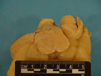
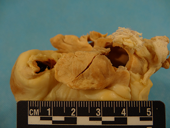
The right atrial appendage is generally triangular in shape in the human heart and may be larger or smaller than the left atrial appendage (1).
|
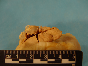
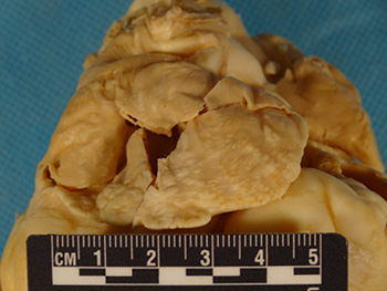
The left atrial appendage is generally tubular in shape in the human heart and may be larger or smaller than the right atrial appendage (1).
|

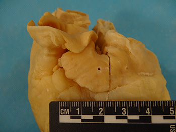
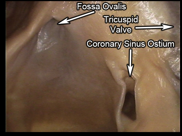
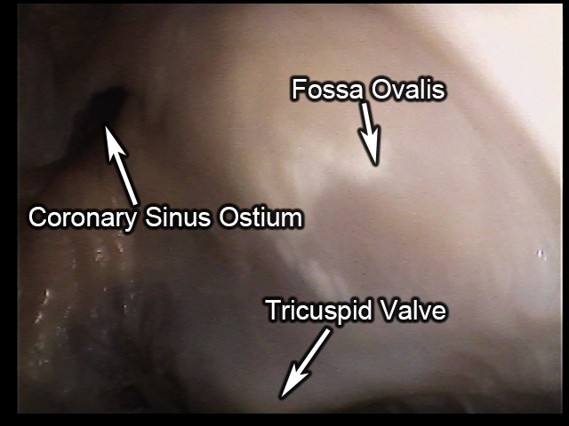
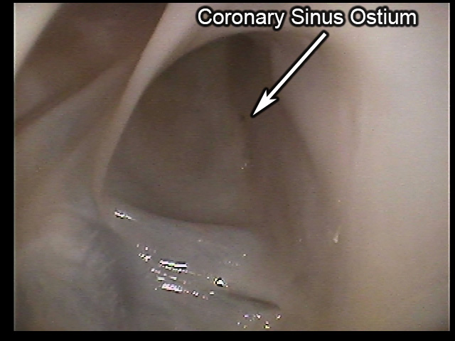
A view of the septal wall within a human right atrium. The coronary sinus ostium of the human heart is partially covered by the Thebesian valve, resulting in a smaller functional ostium than in swine or sheep hearts (1). Not taking the valve into account though, the human heart has a larger superior (postero-lateral) to inferior (antero-septal) coronary sinus ostium diameter than the animal hearts here and a larger lateral (inferior) to septal (superior) diameter compared to the hearts of dogs (1).
|
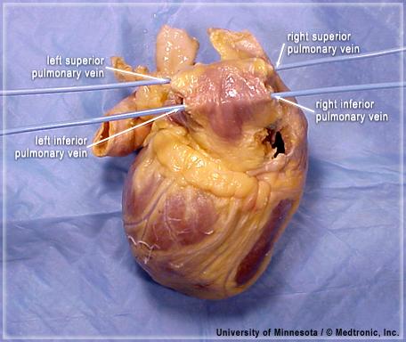
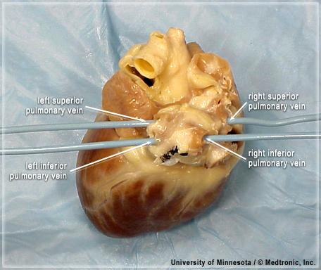
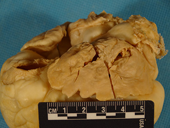
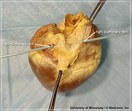
The human heart has 4 or 5 pulmonary veins returning blood from the lungs to the left atrium (1). In this heart, there are 4 pulmonary veins.
|
Canine
In the canine heart, both the right and left atrial appendages take a
tubular shape and the right is generally larger than, or the same size as, the
left (1). Unlike in the human heart, the ostia of the vena cava enter the heart
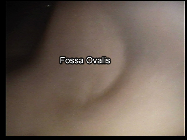
perpendicular to one another (8). The fossa ovalis is positioned much more
posteriorly (caudally) when compared to humans, a trait that is also common to
the hearts of sheep (3). Canine hearts have numerous pulmonary veins, ranging
from 4 to 8, returning blood from the lungs to the left atrium (1). Similar to
human hearts, the left azygous vein is not present in the canine heart (1).
Unlike the human heart, the Thebesian valve does not cover any part of the
coronary sinus ostium (1). Despite this, the functional ostium of the canine
coronary sinus remains similar in size to humans. Likewise, both the lateral
(inferior) to septal (superior) diameter and the superior (postero-lateral) to
inferior (antero-septal) diameter are smaller compared to humans when the
Thebesian valve is not accounted for. The lateral (inferior) to septal
(superior) diameter of the coronary sinus is also significantly smaller in the
canine heart than in both the swine and ovine heart (1).
The right atrial appendage is generally tubular in the canine heart and is larger than or similar in size to the left atrial appendage (1).
|
The left atrial appendage is usually tubular in the canine heart and may be smaller than or similar in size to the right atrial appendage (1).
|
Anywhere from 4 to 8 pulmonary veins may be present in the hearts of dogs to return blood to the left atrium from the lungs (1). In this particular heart, 4 pulmonary veins are present.
|
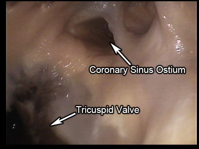
An internal view of the septal wall within a canine right atrium. The coronary sinus ostium of the canine heart is not covered by the Thebesian valve like in the human heart (1). Despite this, the ostium is generally smaller compared to the ostiums of humans, dogs or sheep. The fossa ovalis of the canine heart is more posteriorly (caudally) positioned compared to the fossa ovalis of humans (3).
|
Ovine
In the ovine heart, the right atrial appendage typically has a half-moon
shape and is larger while the left is triangular and smaller (1). As is common
in quadrupeds, the ostia of the vena cava enter the right atrium perpendicularly
(8). The left azygous vein is present in ovine hearts as a tributary to the
coronary sinus, delivering blood directly from the body to the heart (1, 3).
Relative to the human heart, the fossa ovalis of the ovine heart is much more
posteriorly positioned, similar to that seen in canines (3). Similar to the
human heart, the ovine heart has 2 to 4 pulmonary veins returning blood to the
left atrium from the lungs (1). Unlike in humans, the Thebesian valve does not
cover part of the coronary sinus ostium resulting in a larger functional
coronary sinus ostium compared to humans (1). Not taking the valve into account
though, the coronary sinus ostium of the ovine heart has a significantly smaller
superior (postero-lateral) to inferior (antero-septal) diameter than in humans
but similar lateral (inferior) to septal (superior) diameter (1). Compared to
the other animals here, the coronary sinus ostium of the ovine heart is similar
in size to that of swine and large than in canines.
The right atrial appendage is generally half-moon in shape in the hearts of sheep and is larger than the left atrial appendage (1).
|
The left atrial appendage is generally triangular in shape in the hearts of sheep and is smaller than the right atrial appendage (1).
|
The fossa ovalis of the ovine heart is more posteriorly positioned compared to the fossa ovalis of the human heart (3).
|
The coronary sinus ostium of the ovine heart is not covered by the Thebesian valve as it is in humans, resulting in a larger functional diameter (1). Compared to swine, the ostium of the ovine heart is similar in size. Compared to dogs, the ostium is larger (1).
|
Porcine
In the porcine heart, the right atrial appendage typically has a
half-moon shape and is smaller while the left is usually triangular and larger
(1). Unlike in the human heart, the vena cava enter the right atrium
perpendicular to each other. The left azygous vein is present as a tributary to
the coronary sinus, returning blood directly from the body to the heart (1, 8).
Compared to humans, the fossa ovalis is both deeper and more superiorly
positioned in the porcine heart (8). The porcine heart typically has 2 pulmonary
veins returning to the left atrium (1). Like the sheep heart, the Thebesian
valve does not cover part of the coronary sinus ostium resulting in a larger
functional coronary sinus ostium than in humans (1). However, not taking the
valve into account, the superior (postero-lateral) to inferior (antero-septal)
diameter of the coronary sinus ostium is large in humans than in swine but the
lateral (inferior) to septal (superior) diameter is similar in measure. Compared
to ovine hearts, these dimensions are all similar (1). Compared to canine
hearts, the superior to inferior diameter is similar but the lateral to septal
diameter is larger.
The right atrial appendage is usually half-moon in shape in the hearts of swine and is generally smaller than the left atrial appendage (1).
|
The left atrial appendage is generally triangular in shape in the hearts of swine and is usually larger than the right atrial appendage (1).
|
The hearts of swine have two pulmonary veins returning oxygenated blood from the lungs to the left atrium of the heart (1).
|
The coronary sinus ostium of the swine heart is not covered by the Thebesian valve resulting in a larger functional ostium diameter compared to humans (1). Not taking the valve into account, the ostium is smaller in diameter than in the human heart. Compared to the other animals, the coronary sinus ostium is similar in size to that of the ovine heart and larger than in the canine heart (1).
|
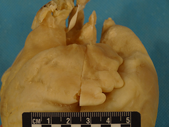
A swine fossa ovalis, located on the septal wall within the right atrium, is pictured here. The porcine fossa ovalis is both deeper and more superiorly positioned compared to the fossa ovalis of human hearts.
|
|
|


