|
 When we receive fresh hearts for research, we attempt to
perfusion fix each heart so that they are preserved in an
end-diastolic state, which is the state when the heart is filled
with blood immediately before contraction. To initiate the
specimen preservation process, four of the great vessels are
cannulated (tubes placed inside) so that there is a cannula
connected to each chamber of the heart. The remaining vessels are
plugged or closed. The heart is then placed in the formalin-filled
lower chamber and its cannulated vessels are connected to the
upper formalin-filled chamber as seen in the figure to the right. The upper
chamber is set to elicit a head pressure of approximately 50 mmHg.
In this set-up, formalin flows antegrade through the aorta into
the coronary arteries. As a result, the formalin perfuses through
the specimen's tissue. The 10% buffered formalin solution
preserves these specimens by cross-linking proteins within the
tissue. After at least 24 hours of perfusion fixation, the heart
is fixed, removed, and stored in an airtight container within
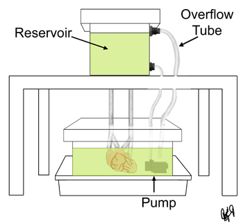
formalin. When we receive fresh hearts for research, we attempt to
perfusion fix each heart so that they are preserved in an
end-diastolic state, which is the state when the heart is filled
with blood immediately before contraction. To initiate the
specimen preservation process, four of the great vessels are
cannulated (tubes placed inside) so that there is a cannula
connected to each chamber of the heart. The remaining vessels are
plugged or closed. The heart is then placed in the formalin-filled
lower chamber and its cannulated vessels are connected to the
upper formalin-filled chamber as seen in the figure to the right. The upper
chamber is set to elicit a head pressure of approximately 50 mmHg.
In this set-up, formalin flows antegrade through the aorta into
the coronary arteries. As a result, the formalin perfuses through
the specimen's tissue. The 10% buffered formalin solution
preserves these specimens by cross-linking proteins within the
tissue. After at least 24 hours of perfusion fixation, the heart
is fixed, removed, and stored in an airtight container within
formalin.
|


