The Visible Heart® is a live beating heart functioning outside of
the body under simulated physiologic conditions. Standard cardiac transplant procedures are employed to arrest a donor heart and prepare it for
reanimation. As with clinical heart recoveries, during the time required to remove the heart and
transplant it into the recipient, the heart remains inactive and is inhibited from spontaneously
contracting. This is accomplished by cooling the heart in an ice slurry (hypothermia) and infusing a
cold high potassium solution cardioplegia through the coronary arteries.
These same conditions or experimental modifications are employed and analyzed for effective need in
our laboratory studies.
The isolated heart apparatus is an
experimental simulation of the donor recipient, providing oxygen and metabolic substrates for the
heart to survive. However, in order to view the internal structures of the beating heart,
non-transparent blood is replaced by a clear synthetic blood-like solution (which does not contain
red blood cells.)
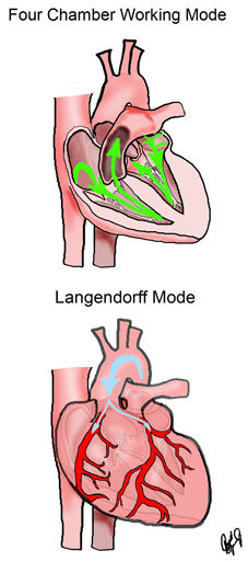
As the coronary system of the heart, which enables the transport of
oxygen and metabolic substrates to the heart, relies on the pressure created by the heart itself for
flow, initially the heart must be externally assisted after transplantation. This is accomplished by
pumping fluid directly into the coronary system and removing fluid from the heart’s chambers to
ease the burden, a technique called the Langendorff mode of perfusion.
Once the heart is capable of maintaining pressures and flows independently, it is weaned (a slow
transition) out of Langendorff mode and the native flow pattern is re-established. Now the heart has
fluid movement through all four chambers and is responsible for the work required to maintain the
flow through the coronary system, hence the name Four Chamber Working
Mode.
This preparation was reviewed and approved by the University of
Minnesota Animal Use and Care Committee. All animals received humane care in compliance with the
“Guide for the Care and Use of Laboratory Animals” published by the National Institutes of
Health. Similarly, the use of the isolated human hearts has be reviewed and approved by the
University of Minnesota human subjects committee.
Human hearts are obtained as generous gifts from LifeSource Organ and Tissue Donation, Inc,
St. Paul, Minnesota. This research is made possible due to the generous gifts of individuals whose
hearts have been donated for research purposes. Their final act of generosity will enhance
understanding of the inner workings of the human heart and contribute to lifesaving advances in
cardiac medicine.
|


