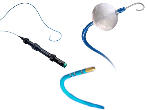
Please select a heart to view
Catheter-CS-1 6250C.mp4 Delivery Systems - Porcine heartShown here is a close-up videoscopic view of a delivery catheter for left-heart transvenous leads being manipulated within the coronary sinus (CS).
Catheter-CS-2 6250S.mp4 Delivery Systems - Porcine heart
Shown here is a close-up videoscopic view of a delivery catheter for left-heart transvenous leads being manipulated within the coronary sinus (CS).
Catheter-steerable-CS Prevail.mp4 Delivery Systems - Porcine heart
Shown here is a videoscopic view of a steerable delivery catheter for left-heart transvenous leads; it is being manipulated within the coronary sinus (CS).
Multi-map-RA ep4rap.mp4 Delivery Systems - Porcine heart
Electrophysiologic studies require the careful placement of diagnostic electrode catheters within the heart. The use of fluoroscopy and intracardiac EMG recordings verifies the position of the electrode catheters.
Three approaches are generally used for advancing the electrode catheter to the heart: 1) femoral venous approach using the inferior vena cava (IVC); 2) subclavian, jugular, or brachial (antecubital) venous approach via the superior vena cave (SVC); or 3) femoral arterial or retrograde approach via the femoral artery.
The approach chosen depends upon the targeted area within the heart, the number of electrode catheters used during the electrophysiologic study, and physician experience and preference. Typically, the IVC and SVC approaches are used for RA, RV, His bundle, and CS electrode catheters.
Shown here is a videoscopic view of four multi-ring mapping/ablation catheters placed into the right heart via the inferior vena cava. These catheters were positioned in the high right atrium, the right ventricular apex, near the His bundle, and within the coronary sinus. This catheter placement is typical during a complex mapping procedure.
Sub-select-catheter-lead-vein-1 6238TEL.mp4 Delivery Systems - Porcine heart
Shown here is an external view of a pre-shaped catheter being advanced into the left anterior descending vein to eventually sub-select a side branch for lead placement.
Sub-select-catheter-lead-vein-2 6248DEL.mp4 Delivery Systems - Porcine heart
Shown here is an external view of a pre-shaped catheter and a flexible tipped intercatheter being advanced into the left anterior descending vein, and then eventually used to sub-select a side branch for lead placement.
Transseptal eptransp.mp4 Delivery Systems - Porcine heart
Electrophysiologic studies require the careful placement of diagnostic electrode catheters within the heart. The use of fluoroscopy and intracardiac EMG recordings verifies the position of the electrode catheters.
Three approaches are generally used for advancing the electrode catheter to the heart: 1) femoral venous approach using the inferior vena cava (IVC); 2) subclavian, jugular, or brachial (antecubital) venous approach via the superior vena cave (SVC); or 3) femoral arterial or retrograde approach via the femoral artery.
The approach chosen depends upon the targeted area within the heart, the number of electrode catheters used during the electrophysiologic study, and physician experience and preference. The femoral arterial approach may also be used for gaining access to the left ventricle and/or left atrium.
As an extension of the IVC approach, an ablation catheter may also be advanced from the right atrium, across the interatrial septum, and into the left atrium using the transseptal approach.
During this approach, the interatrial septum must be punctured before a catheter can be advanced to the left atrium. To create an opening, a transseptal needle is advanced through a long sheath positioned at the foramen ovalis, a thin area of tissue in the interatrial septum. When the needle is withdrawn from the sheath, an ablation catheter is advanced through the septum to the left atrium. At this point the patient must be anticoagulated.
The ablation catheter may then be positioned for mapping and ablation. This approach is useful for left free-wall accessory pathway and AV nodal ablation (atrial fibrillation), as well as future indications for left atrial and left ventricular procedures (e.g., Wolf-Parkinson-White syndrome).
Shown here is a videoscopic view of the transseptal catheterization procedure to access the left atrium through the foramen ovalis (a sheath after a needle puncture).
Transseptal-LA-LV-map eptrablp.mp4 Delivery Systems - Porcine heart
Electrophysiologic studies require the careful placement of diagnostic electrode catheters within the heart. The use of fluoroscopy and intracardiac EMG recordings verifies the position of the electrode catheters.
Three approaches are generally used for advancing the electrode catheter to the heart: 1) femoral venous approach using the inferior vena cava (IVC); 2) subclavian, jugular, or brachial (antecubital) venous approach via the superior vena cave (SVC); or 3) femoral arterial or retrograde approach via the femoral artery. As an extension of the IVC approach, an ablation catheter may also be advanced from the right atrium, across the interatrial septum, and into the left atrium using the transseptal approach.
During this approach, the interatrial septum must be punctured before a catheter can be advanced to the left atrium. To create an opening, a transseptal needle is advanced through a long sheath positioned at the foramen ovalis, a thin area of tissue in the interatrial septum. When the needle is withdrawn from the sheath, an ablation catheter is advanced through the septum to the left atrium. At this point the patient must be anticoagulated. The ablation catheter may then be positioned for mapping and ablation. This approach is useful for left free-wall accessory pathway and AV nodal ablation (atrial fibrillation) as well as future indications for left atrial and left ventricular procedures (e.g., Wolf-Parkinson-White syndrome).
Shown here is a videoscopic view of the transseptal catheterization procedure to access the left atrium through the foramen ovalis. A sheath is placed and then a mapping/ablation catheter is positioned both within the left atrium and ventricle by passing it through the mitral valve.