University of Minnesota
http://www.umn.edu/
612-625-5000
Home
Make a Gift
VHLabs
 
HeartDatabase
 
Right Atrium
Right Ventricle
Pulmonary Trunk
Left Atrium
Left Ventricle
Aorta
Coronary Arteries
Cardiac Veins
External Images
MRI Images
Comparative Imaging
3D Modeling
Plastinates
 
Anatomy Tutorial
Cardiovascular Magnetic Resonance Tutorial
Comparative Anatomy Tutorial
Conduction System Tutorial
Congenital Defects Tutorial
Coronary System Tutorial
Device Tutorial
Echocardiography Tutorial
Physiology Tutorial
 
Project Methodologies
Cardiovascular Devices and Techniques at U of Minnesota
Acknowledgements
References and Links
Atlas in the media
 
Surgery Department
Principal


Anatomy Tutorial
Anatomic Position Attitudinally Correct Anatomy Left Anterior Oblique Anterior Posterior Posterior Fresh Cadaver Dissection

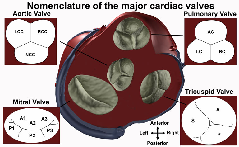
The aortic valve separates the left ventricular outflow tract from the ascending aorta. The aortic valve has also been called the left semilunar valve and the left arterial valve and has three leaflets, or cusps: the left coronary cusp, the right coronary cusp, and the non-coronary cusp. The inlets to the coronary artery system can be found within the sinus of Valsalva, superior to the the leaflet attachments and inferior to the sinotubular junction. The left coronary ostium is found midway between the commissures of the left coronary cusp, and almost immediately branches into the anterior interventricular branch and the circumflex branch. The right coronary ostium is found above the right coronary cusp and gives rise to the right coronary artery. The final cusp is named the non-coronary cusp and is positioned posteriorly relative to the other two cusps.

The mitral valve is also called the bicuspid valve and the left atrioventricular valve. As the name bicuspid valve may suggest, the mitral valve is considered to have two primary leaflets: the anterior and posterior leaflets. The anterior leaflet has also been called the septal, medial, or aortic leaflet, while the posterior leaflet is also referred to as the lateral, marginal, or mural leaflet. Each leaflet is then further broken down into scallops divided by commissures, or zones of apposition. Due to the high variability of leaflet and scallop anatomy, and an alphanumeric nomenclature has been proposed by Carpentier that breaks the leaflets into regions. Three regions are found on the anterior leaflet (A1-A3) with opposing regions on the posterior leaflet (P1-P3). The subvalvular apparatus of the mitral valve consists of chordae tendinae attaching to the anterior and posterior papillary muscles of the left ventricle.

Valve Nomenclature

The relative positions of the aortic, mitral, pulmonary, and tricuspid valves are shown in the diagram of the heart at the center of the figure. The aortic valve has three cusps: the left coronary cusp (LCC), the right coronary cusp (RCC), and the non-coronary cusp (NCC). The mitral valve has an alphanumeric nomenclature that numbers from the anterior to the posterior, with respect to the heart, and attaches an A or a P in front of the anterior or posterior leaflets, respectively (A1-A3, P1-P3). The pulmonary valve has three cusps: the anterior cusp (AC), the left cusp (LC), and the right cusp (RC). The tricuspid valve has three leaflets named the anterior (A), septal (S), and posterior (P).

The pulmonary valve separates the right ventricular outflow tract of the right ventricle from the pulmonary trunk. The pulmonary valve can also be referred to as the pulmonic valve, the right semilunar valve, and the right arterial valve. Its three leaflets, or cusps, are difficult to name because of the oblique angle of the valve. Its nomenclature is therefore derived based on the nomenclature of the aortic valve, which lies in proximity to it. The two leaflets attached to the septum are named the left and right leaflets, and correspond to the right and left leaflets of the aortic valve, which they face. The third leaflet is called the anterior leaflet or the non-coronary leaflet (to maintain the nomenclature of the aortic valve).

The tricuspid valve, also called the right atrioventricular valve, gets its name because it is generally considered to have three leaflets: the anterior, posterior and septal leaflets. Of these, the anterior, also called the infundibular or anterosuperior, leaflet is typically the largest. The posterior leaflet is also referred to as the inferior or marginal leaflet and the septal leaflet is also referred to as the medial leaflet. Terminating on the ventricular side of the tricuspid valve leaflets, the chordae tendinae are connected to three papillary muscles in the right ventricle. In humans, the three papillary muscles of the right ventricle have highly variable anatomy. The anterior papillary muscle is usually the most prominent, with the moderator band terminating at its head. The moderator band typically originates from the septal papillary muscle. The septal papillary muscle is normally the least prominent, and is missing 21.4% of the time.


© 2021 Regents of the University of Minnesota. All rights reserved. The University of Minnesota is an equal opportunity educator and employer. Privacy Statement