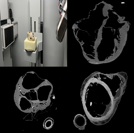
This section contains micron-resolution Computed Tomography (microCT) images of isolated human hearts.
Prior to imaging, the great vessels were cannulated and the hearts perfusion fixed in 10% buffered formalin for a period of 24-48 hours under 40-50 mmHg such that the hearts would retain an end-diastolic shape. After perfusion fixation, the hearts were filled with shaving cream prior to provide gentle support and hydration for thin structures during scanning. 360 degree radiographic images were acquired for each heart using a North Star Imaging x3000 industrial CT scanner. Images were reconstructed into CT datasets using the efX-CT software provided with the CT scanner. These datasets use isovolumetric resolutions, meaning that each pixel's width and height are the same, and this is the same size as the slice thickness. Each data set will have its own resolution based on the specific scan parameters used at the time of scanning. Resolutions will be in microns.
Some hearts were stained with Iodine prior to scanning in order to highlight the fiber structures and cardiac conduction system of the heart.