|
MRI Preparation
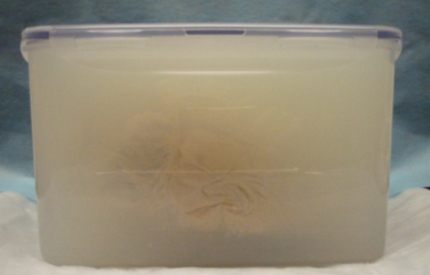
Hearts (saturated with formalin) were placed in 12" x 7" x 6" sealable polymer container and submerged in 0.7% agar gel at approximately 45°C. The hearts were then palpated to ensure that all possible air are ejected from the chambers and major vessels, reducing any air-tissue susceptibility artifacts, before being suspended in an anatomically correct position and allowing the gel to cool and set, as seen in the figure to the right. Once cooled to room temperature (~23°C), all supports used to suspend the heart are removed and any remaining air volume within the container was displaced with gel. Then each are brought to an MRI scanner and images are obtained so to create both still images and imaging stacks, including: a short axis valve, a short axis ventricle, a 4 chamber, a 2 chamber left, a 2 chamber right, a short axis stack, a 4 chamber stack and a 2 chamber stack.
After imaging, the hearts are then removed from the agar gel and rinsed thoroughly in warm water, approximately 45°C. In order to preserve the anatomy of the specimens, the majority of the gel was removed in an antegrade manner through the pulmonary trunk and the aorta, and subsequent cannulation of the superior vena cava and a pulmonary vein and circulation of warm water through the heart was used to dissolve any remaining gel.
|