|
The conduction systems of the hearts of large mammals are very similar (1). The
main structures of the conduction system include the sinoatrial node (SA node),
the atrioventriular node (AV node), the bundle of His, the right and left main
bundle branches and the Purkinje fibers; all of these structures are present in
humans, canine, swine and ovine (1). Pacemaking is performed by the SA node,
located high on the right atrial wall near the junction of the superior vena
cava and the right atrium (3, 10, 11). Beginning in the SA node, the
depolarization of cells, which triggers contraction, travels through the atria
to the AV node, located subendocardially in the area between the coronary sinus
ostium, membranous septum and the septal/posterior commissure of the tricuspid
valve know as the "triangle of Koch". The signal then spreads to the bundle of
His, also located in the triangle of Koch, which penetrates through the central
fibrous body separating the atria and ventricles. The bundle of His then
bifurcates into right and left main bundle branches, which branch further to
become Purkinje fibers that spread conduction to the ventricles. Differences in
the conduction system primarily reside in the arrangement of the transitional
and compact components of the AV node and in the length and route of the bundle
of His.
Human
The AV node of the human heart is located at the base of the atrial septum,
anterior to the coronary sinus and just above the tricuspid valve, a location
that is similar to that seen in dogs (27). Unlike in dogs and sheep, the
transition point, where the AV node meets the bundle of His, is difficult to
distinguish. The bundle of His is located just below the membranous septum at
the crest of the interventricular septum (27). The unbranched portion of the
bundle of His extends 2 to 3 millimeters before penetrating the central fibrous
body for a length of 0.25 to 0.75 millimeters. The bundle then bifurcates
immediately after emerging from the central fibrous body (27).
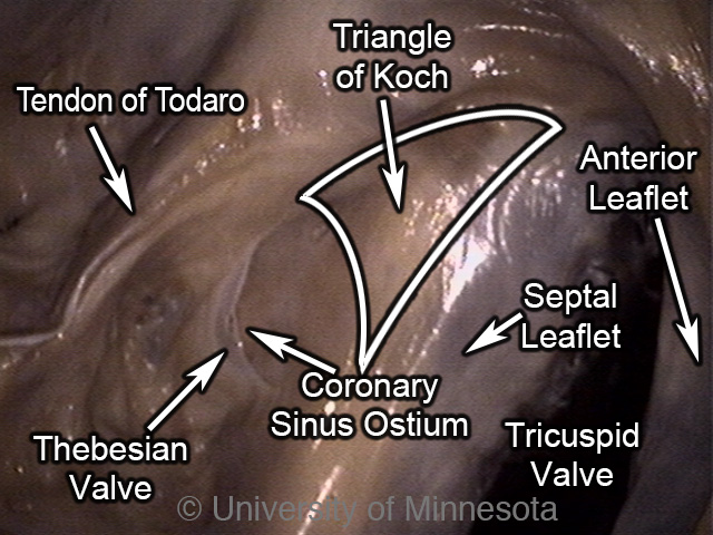
An internal image of the septal wall within a human right atrium. Visible is the triangle of Koch where the AV node and bundle of His reside subendocardially.
|
Canine
The AV node of the canine heart is located at the base of the atrial septum,
anterior to the coronary sinus and just above the tricuspid valve, in a position
similar to that seen in humans (30). The junction joining the AV node and the
His bundle consists of internodal tracts of myocardial fibers. At least three
different His bundle branches extend from the AV node via a proximal branch of
the His bundle (30). The penetrating bundle of the bundle of His is 1 to 1.5 mm
long, significantly longer than that of humans, and runs forward and downward,
just beneath the endocardium, through the fibrous base of the heart (30).
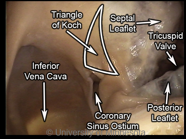
An internal image of the septal wall within a canine right atrium. Visible is the triangle of Koch where the AV node and bundle of His reside subendocardially.
|
Ovine
The AV node of the ovine heart is located at the base of the atrial septum,
anterior to the coronary sinus and just above the tricuspid valve (29). This
position is also at the junction of the middle and posterior one-third of the os
cordis. The junction between the AV node and His bundle is clearly defined as
finger-like projects where the two tissues overlap. These two tissue types are
easily identifiable by both size and histological examinations (29). The
unbranched portion of the His bundle passes beneath the os cordis to reach the
right side of the ventricular septum and remains relatively deep thereafter. The
portion passing through the central fibrous body is ~1 mm long and extend 4 to 6
mm beyond the central fibrous body before bifurcating, a much more anterior
bifurcation than that seen in humans (29).
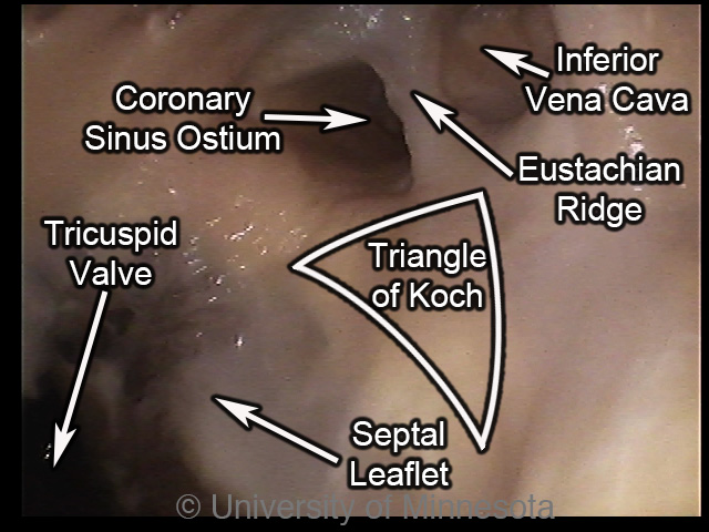
An internal image of the septal wall within a ovine right atrium. Visible is the triangle of Koch where the AV node and bundle of His reside subendocardially.
|
Porcine
The AV node of the porcine heart lies on the right side of the crest of the
ventricular septum, more inferiorly on the septum than in humans (28). From the
bundle of His, the conduction path climbs to the right side of the summit of the
ventricular septum and enters the central fibrous body. Notably, the bundle of
His bifurcates much earlier along the conduction pathway compared to the
conduction system of humans (28).
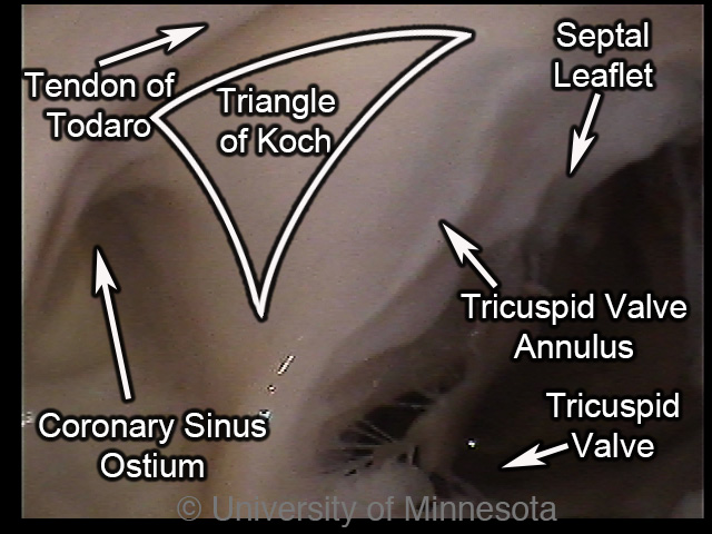
An internal image of the septal wall within a swine right atrium. Visible is the triangle of Koch where the AV node and bundle of His reside subendocardially.
|
|


